세극등현미경에서 보존액에 담긴 기증각막을 관찰할 수 있는 장치의 개발
Development of an Instrument for Slit-lamp Examination of Donor Corneas in Preservation Medium
Article information
Abstract
목적
각막이식수술 전 정확한 이식 적합성 평가를 위하여 기증각막이 담긴 보존액 병을 고정시킬 수 있는 장치를 개발하였으며, 국내 기증각막을 대상으로 세극등현미경검사를 시행하여 고안된 장치의 효용성을 평가해보고자 한다.
대상과 방법
2023년 2월부터 2023년 3월까지 여의도성모병원에서 기증받는 국내 기증각막 2안을 대상으로 하였다. 기증각막이 담긴 보존액 병을 고안된 장치의 홀더에 장착하여 세극등현미경에 설치하면, 세극등현미경의 빔은 장치의 거울에 의해 반사되어 보존액 내의 각막에 입사하고, 이를 세극등현미경에서 관찰할 수 있다. 각막내피가 보존액 병의 위를 향한 상태와 바닥을 향한 상태에서 세극등현미경검사를 시행하고 전안부 사진을 촬영하였다. 또한, 고안된 장치를 이용하여 기증각막에 경면현미경검사와 전안부 빛간섭단층촬영을 시행하였다.
결과
기증각막이 담겨있는 보존액 병을 장치에 고정한 상태에서 세극등현미경을 이용하여 공막 변연부를 360° 관찰하여 버트닝 상태를 확인하고, 세극등현미경의 좁은 빔으로는 각막 단면을 확인할 수 있었다. 역조명법을 이용하여 각막상피세포를 관찰할 수 있었으며, 경면반사법을 통해 내피세포층이 확인되었다. 그러나 경면현미경검사 및 빛간섭단층촬영검사 시 내피세포밀도와 각막두께는 측정되지 않았다.
결론
저자들이 고안한 기증각막검사용 장치는 국내 기증각막에 대한 세극등현미경검사를 쉽고 안정적으로 시행할 수 있게 하며, 각막내피나 두께에 대한 정성적인 평가가 가능하였다.
Trans Abstract
Purpose
To evaluate the effectiveness of an instrument devised for slit-lamp examination of donor corneas suspended in preservation medium.
Methods
The study examined two donor corneas received at Yeouido St. Mary's Hospital in February 2023 and March 2023. The instrument has three main components: a plastic holder to hold the preservation medium bottle, a cube with a mirror for reflecting the slit beam, and a stand to attach the device to the slit-lamp. Using the instrument, the donor corneas were examined via slit-lamp: microscopy with the endothelium facing upward and downward. Specular microscopy and anterior segment optical coherence tomography (OCT) were also performed on the preserved donor corneas.
Results
Slit-lamp examination of donor corneas in preservation medium using the instrument showed overall corneal buttoning and optical sections of the donor cornea. Using specular reflection and retroillumination, the endothelial layer was partially visible. However, specular microscopy and anterior segment OCT could not examine the donor cornea in preservation medium using the instrument.
Conclusions
The devised instrument facilitates slit-lamp examination of donor corneas in preservation medium, enabling a qualitative assessment of donor corneas before corneal transplantation surgery.
각막이식수술은 각막에 기능적으로나 구조적으로 회복 불가능한 손상이 발생하여 시력이 저하된 환자의 각막을 제거하고 공여자로부터 기증받은 투명한 각막을 이식하는 수술이다.1 각막이식 전 기증각막의 상태에 대한 정확한 평가는 수술의 예후에 영향을 미치는 요소 중 가장 중요한 요인이다. 기존에 기증각막은 기증자에 대한 임상적 기초 정보와 각막내피세포 및 각막두께 측정, 세극등현미경검사를 통해 이식 적합성을 평가하여 수술을 시행해왔다.2,3
수입 기증각막의 경우에는 각막 보존액이 담겨있는 corneal viewing chamber (Krolman, Boston, MA, USA)에 각막절편이 고정된 상태로 보관이 되어, 세극등현미경을 이용하여 기증각막의 전면 및 후면을 보다 용이하게 관찰할 수 있다(Fig. 1A).4 그러나 국내 기증각막의 경우 각막 보존액(Optisol-GS, Bausch & Lomb Inc., Rochester, NY, USA)에 별도의 고정장치 없이 담긴 상태로 보관하여, 각막이 고정되어 있지는 않기 때문에 세극등현미경으로 관찰이 어렵고, 대신에 병을 들고 각막을 육안으로밖에 관찰할 수 없다(Fig. 1B).
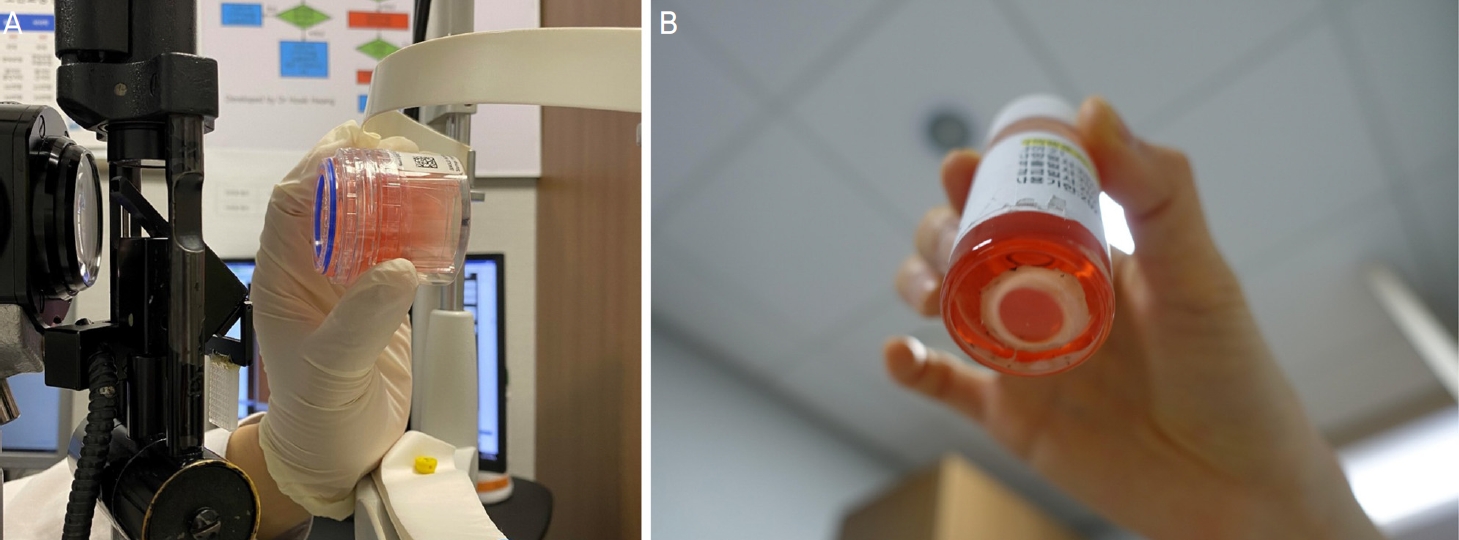
Comparison between an imported donor cornea stored in Krolman corneal viewing chamber and a domestic donor cornea stored in the preservation medium bottle. (A) Slit-lamp examination of an imported donor cornea is facilitated by the Krolman viewing chamber. (B) Because a domestic donor cornea suspended in preservation medium cannot be held in proper position for slit-lamp examination, inspection is only possible with the naked eye, holding the bottle up.
본 연구에서는 보존액 병에 담긴 국내 기증각막을 세극등현미경으로 쉽게 관찰할 수 있도록 한 장치를 저자들이 개발하였으며, 국내 기증각막의 이식수술 전 검사 시 장치의 효용성을 평가하여 이를 보고하고자 한다.
대상과 방법
2023년 2월부터 2023년 3월까지 여의도성모병원에서 기증받는 국내 기증각막 2안을 대상으로 하였다. 첫 번째 기증각막은 기저 질환으로 고혈압이 있으며 안과적 병력이 없는 52세 뇌사자의 각막이며, 두 번째 기증각막은 내과적, 안과적 병력이 없는 21세의 사후 기증각막이었다. 본 연구는 헬싱키선언(Declaration of Helsinki)을 준수하였으며, 여의도성모병원 임상연구윤리위원회(Institutionary Review Board, IRB)의 심사 후 승인을 받았다(승인번호: 2023-0384-0001). 사전에 직계가족의 장기기증 서면 동의가 시행된 자에 대하여 안구적출을 시행하였으며, 기증받는 병원이나 장기기증자로부터 기증자의 성별/나이, 사인 및 기저 병력(전신 및 안과적 병력), 사망시각 등의 정보를 제공받았다.
먼저 적출된 기증자 안구를 거즈로 감싼 채 손으로 들고 세극등현미경에서 각막상피결손, 기질부종 및 기질혼탁, 노인환, 데스메막 주름과 그 정도를 평가하였다. 각막 버트닝(corneal buttoning) 시 각막윤부로부터 약 3 mm 남도록 공막을 잘라 각공막 이식편을 만들었으며, 각막 보존액인 Optisol-GS (Bausch & Lomb Inc.) 용액에 각막내피가 위로 향하도록 담아 4°C 냉장 상태로 보관하였다. 기증각막이 보존액 병에 담긴 상태에서 서울성모병원 안은행에 있는 기증각막용 경면현미경(Eye Bank Kerato Analyzer; EKA-10, Konan Medical Inc., Hyogo, Japan)으로 내피세포밀도와 각막두께를 측정하였다.
고안된 장치는 세 가지 주요 부분으로 구성되어 있다. 첫 번째로, 보존액 병을 고정할 수 있는 플라스틱 홀더가 있다. 플라스틱 홀더는 직경 30 mm, 길이 116 mm의 플라스틱 원심관 튜브(SPL 50 mL conical tube, SPL Life Sciences, Pocheon, Korea)의 위쪽 25 mm를 잘라내어 사용하였다. 홀더 바로 아래에는 내부에 거울이 장착된 큐브(30 mm Cage Cube-Mounted UV-Enhanced Aluminum Turning Mirror; CCM1-F01/M, Thorlabs, Newton, NJ, USA)가 있으며, 큐브 아래쪽으로 세극등현미경의 턱받침에 고정할 수 있는 거치대가 있다(Fig. 2A). Fig. 2B는 고안된 장치를 세극등현미경에 설치한 모습이다. Fig. 3의 모식도에서처럼 고안된 장치에 입사된 세극등현미경의 빔은 큐브 내의 거울에 의해 반사되어 보존액 내의 각막에 입사한다. 이를 세극등현미경으로 관찰할 수 있다.
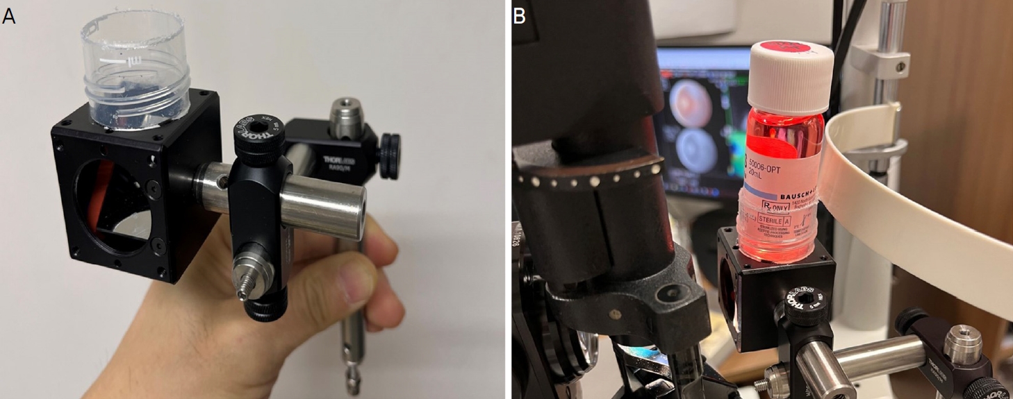
The instrument devised for evaluation of donor corneas in the preservation medium bottle. (A) The instrument consists of three major pieces: a plastic holder for holding the bottle, a cube cage with a mirror placed at a 45° angle, and a metal rod which allows attachment to the slit-lamp. (B) Slit-lamp attachment for the examination of a donor cornea in the preservation medium bottle.
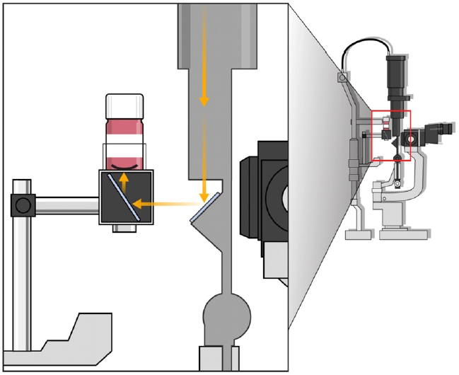
A schematic diagram of slit-lamp examination of a donor cornea in the preservation medium bottle using the instrument. The red box shows the magnified view of a donor cornea within the bottle and its position in relation to the light source (yellow arrows).
먼저, 각막의 내피가 보존액 병의 위쪽으로 향하도록 한 다음 보존액 병을 고안된 장치의 플라스틱 홀더에 장착하면, 검사자는 세극등현미경의 슬릿빔의 각도를 조절하고 세극등현미경을 앞뒤로 움직여서 각막에 초점을 맞춘 뒤, 전안부 사진을 촬영하였다. 세극등의 넓은 빔을 이용하여 병을 360° 돌려가면서 공막 변연부를 관찰하였다. 세극등현미경 빔의 너비를 줄여서 각막의 두께를 확인하였다. 입사각을 거의 0°로 하면 각막을 투과한 빛은 보존액 병의 흰색 뚜껑에서 반사하여 역조명법과 같은 효과를 갖게 된다. 이를 통해 각막상피세포를 관찰하였다. 데스메막 주름이 있는 부분에 대해서 경면반사법을 이용하여 내피세포를 촬영하였다.
다음으로, 각막의 내피가 보존액 병의 바닥 쪽으로 향하도록 한 다음 병을 장치에 고정한 상태에서 세극등현미경검사를 시행하여 포도막 조직이 잘 박리되었는지 확인하고, 경면반사법을 이용하여 내피세포를 관찰하였다.
고안된 장치를 이용하여 여의도성모병원에서 통상적인 진료에 사용되는 경면현미경(CellChek 20 Specular Microscope, Konan Medical, Irvine, CA, USA)을 통한 기증각막의 내피세포 촬영을 시행하였으며, 전안부 빛간섭단층촬영검사(Topcon DRI OCT Triton Swept source OCT, Topcon, Tokyo, Japan)를 실시하여 각막두께를 측정해 보았다(Fig. 4).
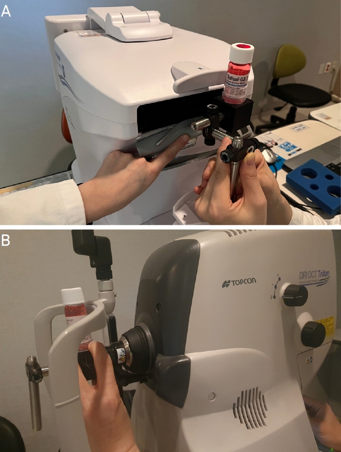
Donor corneal tissue analyses using the instrument. (A) Specular microscopy examination (CellChek 20 Specular Microscope, Konan Medical, Irvine, CA, USA) and (B) anterior segment optical coherence tomography scan (Topcon DRI OCT Triton, Topcon, Tokyo, Japan) of the cornea from the 52-year-old donor using the instrument.
결 과
세극등현미경검사
각막내피세포가 위를 향하도록 한 상태에서 세극등현미경의 넓은 빔으로 기증각막의 가장자리를 확인하였다. 52세 기증각막에서 남아있는 공막의 폭을 확인할 수 있었으며 결막 박리가 불완전하다는 것을 확인할 수 있었다(Fig. 5). 군날개나 노인환 등의 각막 주변부 병변이 없다는 것도 알 수 있었다. 보존액 병을 360° 돌리면 기증각막의 공막 변연부의 폭과 결막 박리 상태를 360° 전체적으로 확인할 수 있었다(Supplementary Video 1). 세극등현미경의 얇은 빔으로 저배율 및 고배율의 각막 단면 이미지를 얻어, 각막실질의 전반적인 투명도, 대략적인 각막의 두께를 확인할 수 있었다(Fig. 6).
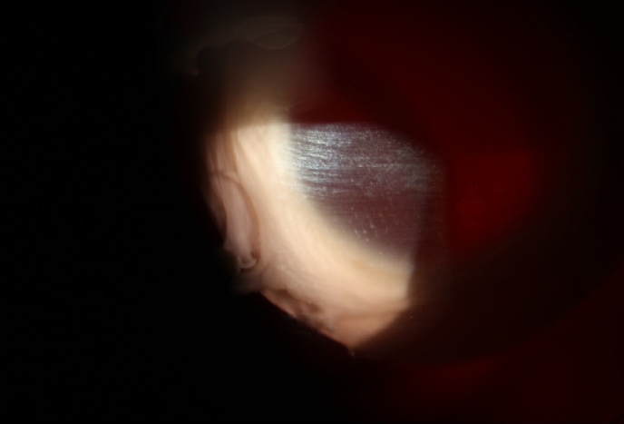
Slit-lamp photograph of the cornea from the 52-yearold donor, showing that the conjunctiva on corneoscleral button is not thoroughly dissected (×10 magnification).
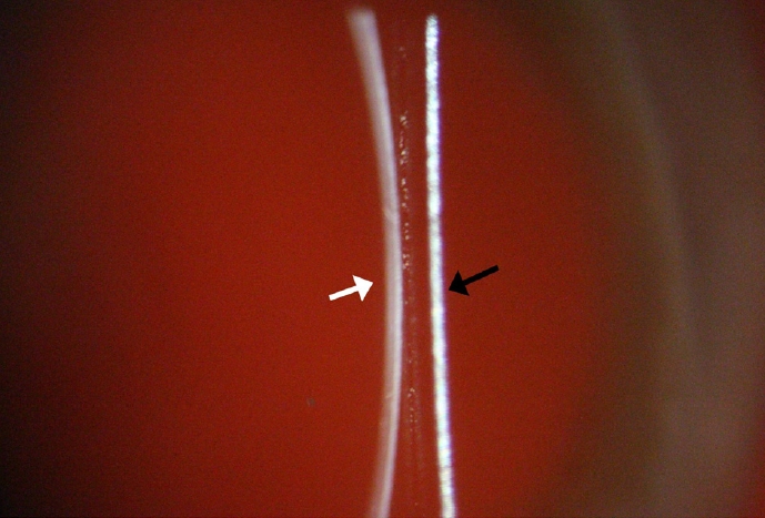
Slit-lamp photograph of the cornea from the 52-yearold donor with a narrow focal slit beam projected at the center of cornea. It is possible to visualize the optical sections of cornea (white arrow) just above the section of the bottom of the preservation medium bottle (black arrow) (×16 magnification).
21세 기증각막에서 역조명법을 이용하였을 때 내피세포층은 관찰할 수 없었고, 상피에 붙어있는 공막조각과 상피세포가 온전하게 붙어있지 않아 불균질하게 보이는 부분에서 각막상피의 결손을 확인할 수 있었다(Fig. 7). 경면반사법에서는 데스메막 주름이 있는 부위에서는 강한 반사 뒤로 내피세포층을 확인할 수 있었다(Fig. 8, Supplementary Video 2).
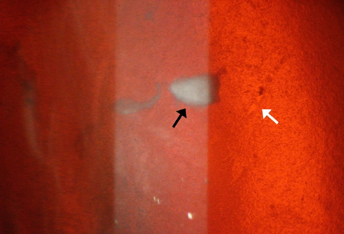
Slit-lamp photograph of the cornea from the 21-yearold donor, placed endothelium facing up. A narrow-moderate width slit beam was projected at a narrow angle (nearly 0°) for retroillumination effect. It is possible to visualize remnant debris from sclera (black arrow) and focal areas of epithelial loss (white arrow) (×40 magnification).
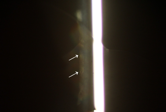
Slit-lamp photograph of the cornea from the 21-yearold donor, placed endothelium facing up. Using specular reflection, endothelial layer is visible at regions of Descemet membrane folds (white arrows) (×40 magnification).
21세 기증각막의 내피가 병의 바닥 쪽으로 향하도록 놓여있는 상태에서는 세극등현미경의 넓은 빔으로 각공막 절편에 포도막 조직이 잘 박리되어 있음을 확인할 수 있었다(Fig. 9). 그리고 경면반사법을 이용하여 내피세포층을 관찰할 수 있었다(Fig. 10).
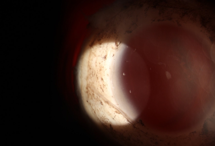
Slit-lamp photograph of the cornea from the 21-yearold donor, placed endothelium facing down. The uvea is completely dissected and removed from corneoscleral button (×10 magnification).
경면현미경검사(specular microscopy)
서울성모병원 안은행에서 기증각막의 검사에 사용되는 기증각막용 경면현미경으로 보존액 병에 담겨있는 동일한 기증각막에 대하여 기증각막 2안 모두에서 내피세포 촬영이 가능하였으며, 내피세포의 밀도와 세포 면적의 변이계수, 세포 모양의 다형성 등을 확인할 수 있었다. 52세 기증각막의 각막내피세포밀도는 2,688/mm2 , 21세 기증각막의 각막내피세포밀도는 3,048/mm2 로 측정되었다(Fig. 11). 반면, 저자들이 고안한 장치를 이용하여 시행한 환자용 비접촉식 경면현미경검사에서는 기증각막 2안 모두에서 내피세포 촬영이 불가능하였다(Fig. 12A).
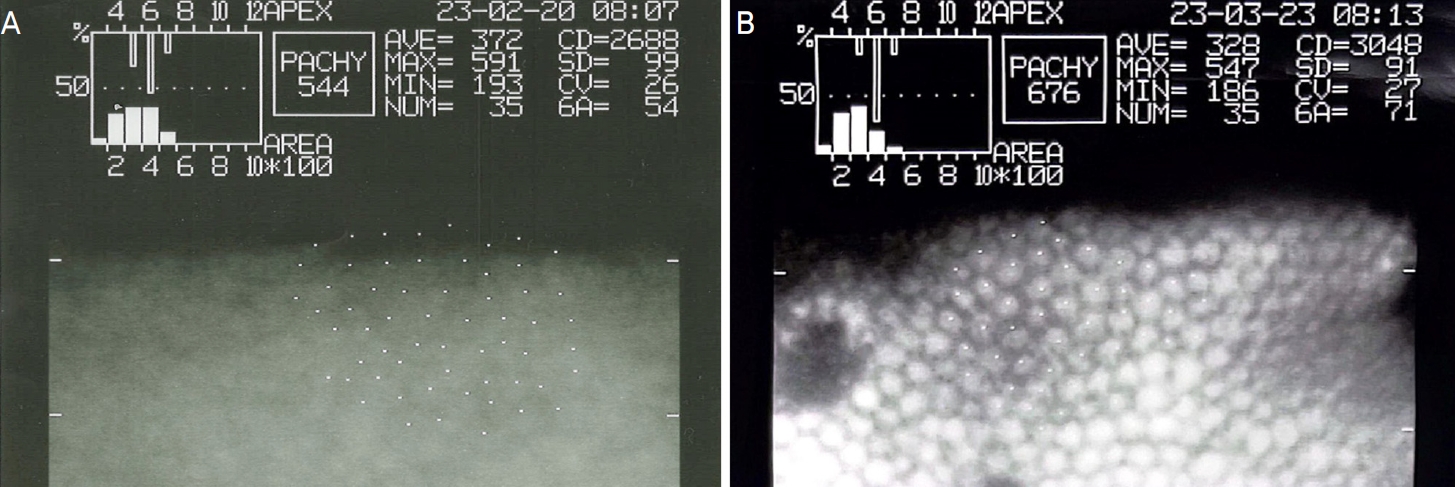
Endothelial cell count reports of donor corneas using the corneal specular microscopy (Eye Bank Kerato Analyzer, EKA-10, Konan Medical Inc., Hyogo, Japan) at Seoul St. Mary’s Hospital. The endothelial cell dencity was estimated (A) 2,688 cells/mm2 in the cornea from the 52-year-old donor and (B) 3,048 cells/mm2 in the cornea from the 21-year-old donor.
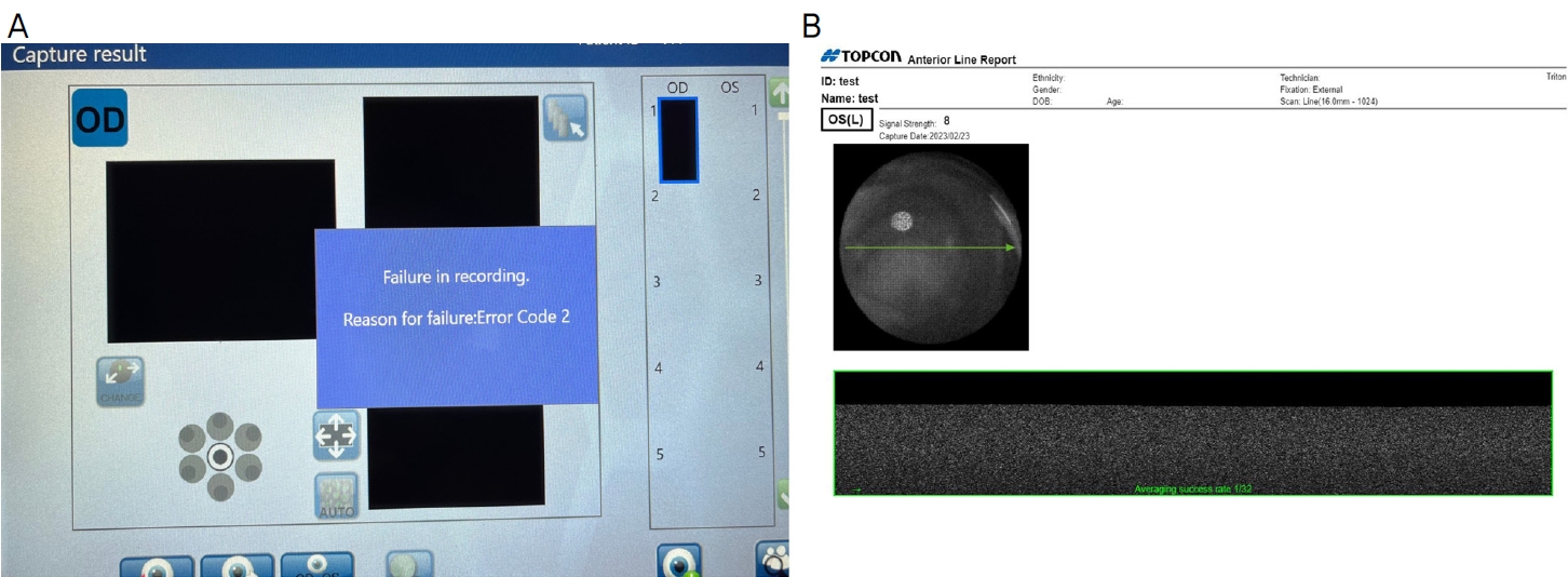
Failed results of donor corneal tissue analyses using the instrument. (A) Corneal specular microscopy (CellChek 20 Specular Microscope, Konan Medical, Irvine, CA, USA) and (B) anterior segment optical coherence tomography scan (Topcon DRI OCT Triton, Topcon, Tokyo, Japan) failed to examine the donor cornea in the preservation medium bottle using the instrument.
전안부 빛간섭단층촬영검사(anterior segment optical coherence tomography [OCT])
고안된 장치를 이용하여 기증각막에 시행한 전안부 빛간섭단층촬영검사에서도 각막이 식별되지 않아 기증각막 2안 모두에서 각막 단층에 대한 촬영이 불가능하였다(Fig. 12B).
고 찰
안은행에서는 수술 전 기증각막의 상태를 파악하기 위해서 세극등현미경검사, 경면현미경검사, 각막두께 측정 등을 검사하고 있다. 세극등현미경검사로는 각막의 혼탁 및 부종, 기질침윤, 상피결손, 신생혈관, 익상편 등의 전반적인 각막 병리 소견에 대해 확인할 수 있으며,5 경면현미경검사는 이식 각막의 생존에 가장 중요한 각막내피세포의 형태와 밀도를 측정하는 데 필수적인 검사이다.6 또한, 각막두께는 각막부종의 여부를 파악하고 각막내피세포의 기능을 간접적으로 알 수 있도록 하는 중요한 검사이다.7
본 연구에서는 보존액 병에 담긴 국내 기증각막에 대하여 세극등현미경검사를 시행하기 어려웠던 한계점을 극복하기 위하여 기증각막검사용 장치를 개발하였으며, 국내 기증각막 2안을 대상으로 세극등현미경검사를 시행하여 고안된 장치의 성능을 확인할 수 있었다. 기증각막이 담겨있는 보존액 병을 장치에 고정한 상태에서 병을 천천히 회전시켜 공막 변연부와 각막윤부를 360° 관찰할 수 있었다. 넓은 빔으로 기증각막을 전반적으로 관찰하여 결막과 포도막 조직의 박리가 잘 되어 있는지 확인하고, 좁은 빔으로는 각막 단면을 확인할 수 있었다. 역조명법을 이용하여 각막상피세포를 관찰할 수 있었으며, 경면반사법을 통해 내피세포층을 확인할 수 있었다.
기증각막의 내피세포 상태는 각막이식수술의 예후에 영향을 미치는 가장 중요한 요인 중 하나로 술 전 내피세포의 형태와 밀도가 중요하다.8 수입 기증각막에 세극등현미경검사를 시행하는 경우에는 각막이 담긴 corneal viewing chamber를 앞뒤로 돌려가며 각막의 내피면과 상피면을 여러 각도에서 동일하게 잘 관찰할 수 있다. 본 연구에서 고안된 장치를 이용하여 국내 기증각막에 검사를 진행하는 경우 각막내피세포를 위로 향하도록 하고 관찰하였을 때는 데스메막 주름이 있어 굴곡진 부분에서는 경면반사법에 의해 각막실질 뒤의 내피세포층을 부분적으로 관찰할 수 있었다. 반면, 각막내피가 아래로 향하도록 하고 관찰하였을 때는 내피세포층을 직접 관찰할 수 있었다.
이번에 개발한 장치를 이용하여 내피세포밀도를 측정할 수 있다면 가장 이상적일 것이다. 그러나 기증각막용 경면현미경을 이용하면 보존액 병에 있는 기증각막의 내피세포 밀도와 형태를 측정할 수 있었던 것에 반해, 고안된 장치에 보존액 병을 장착한 상태에서 환자용 일반 경면현미경으로 검사를 시행하였을 시에는 내피세포를 측정할 수 없었다. 비접촉 경면현미경의 작동 원리는 각막의 입사된 빛의 0.02%가 내피세포층에서 반사되는데 이를 촬영하는 것이다.9 아마도, 이번 장치를 이용할 때는 경면현미경의 빛이 장치의 거울에 반사되어 각막으로 입사하기 때문에 빛의 작동 경로가 너무 길어지면서 반사한 빛을 경면현미경이 인식하지 못하였기 때문으로 생각된다.
기증각막의 두께 측정은 각막의 이식 가능성 및 각막내피세포의 기능을 간접적으로 확인하는 데 꼭 필요하다. 기증각막의 두께 측정은 세극등현미경, 초음파 또는 빛간섭단층촬영으로 가능하다. 기증각막이 보존액 병에 담긴 경우에는 초음파는 사용할 수 없다. 본 연구에서 고안한 장치에 보존액 병을 고정하고 세극등현미경으로 관찰하였을 때는 정성적으로 각막두께를 알 수 있었다. 만약 빛간섭단층촬영이 가능하였다면 각막두께를 정량적으로 측정할 수 있었을 텐데 전안부 빛간섭단층촬영을 이용하여 보존액 병에 담긴 기증각막 촬영은 실패하였다. 고안된 장치를 이용하여 기증각막에 전안부 빛간섭단층촬영 검사를 시행하였을 때 각막이 인식되지 않았는데, 경면현미경검사와 마찬가지로 빛의 작동 경로가 길어져서 측정에 제한이 있는 것으로 생각된다.
저자들은 보존액 병에 담긴 기증각막을 세극등현미경으로 촬영한 전안부 이미지를 이용하여 각막두께를 정량적으로 측정하기 위한 시도를 하였다. 보존액 병 바닥의 두께는 전안부 빛간섭단층촬영검사(Topcon DRI OCT Triton Swept source OCT, Topcon)를 이용하여 측정하였으며, 또한 보존액 병을 파손시켜 바닥의 실제 두께를 버니어 캘리퍼스로 다시 측정하였다. 그 결과 전안부 빛간섭단층촬영검사를 이용하여 측정한 보존액 병 바닥의 두께는 953 µm였으며, 버니어 캘리퍼스로 측정하였 때는 0.95 mm (950 µm)였다. 세극등현미경 촬영 사진에서 각막 단면의 두께와 보존액 병 바닥의 단면의 두께를 Image J (NIH, Washington, USA)로 픽셀 수로 측정하여 비례식을 이용하여 각막두께를 측정하였다. 이렇게 측정한 52세 기증각막의 두께는 1,137.5 µm로, 기증각막 경면현미경검사에서 측정된 기증각막의 두께인 544 µm보다 지나치게 큰 값을 보였다. 이는 세극등현미경의 빔은 평행광선이 아니라 빔이 모였다가 펴지는 모양으로, 보존액 병 바닥에 빔이 초점을 맺으면 보존액 병 바닥과 거리가 떨어져 있는 각막에서는 훨씬 퍼져 있는 빔이 지나가기 때문이며, 이러한 방법으로는 정량적으로 중심각막두께를 측정하는 것은 불가능하다. 다른 방법으로는 중심각막두께 측정을 위해서 Scheimpflug 카메라 방식의 각막지형도 검사계(Pentacam [Oculus, Wetzlar, Germany], Galilei dual Scheimpflug system [Ziemer Ophthalmic Systems AG, Port, Switzerland])를 이용해볼 수도 있다. 이는 추후 연구에서 시도해보고자 한다.
이전에도 해외에서는 각막 보존액 병에 기증각막이 담긴 상태로 세극등현미경에 고정하여 검사를 시행할 수 있는 장치가 개발된 적이 있다.10 그가 개발한 장치는 보존액 병을 끼워 넣을 수 있는 링이 달려있는 황동 막대와 수직 및 회전을 가능하게 하는 클램프 및 나비나사 그리고 45° 각도로 부착된 거울로 구성되어 있다. 거울에 반사된 세극등현미경의 빔은 보존액 병의 바닥으로 입사하여 기증각막에 대한 세극등현미경검사가 가능하게 하였다.10 해외에서 사용하는 corneal viewing chamber는 위와 아래가 투명한 플라스틱 창으로 되어 있으며, 각막을 내피세포가 위로 가도록 chamber 안에 고정하도록 되어 있다. 따라서, 세극등현미경으로 기증각막의 내피면이나 상피면을 잘 관찰할 수 있으며, 기증각막용 경면현미경으로 각막내피세포밀도를 측정하며, 전안부 빛간섭단층촬영검사로 중심각막두께를 측정할 수 있다.11 국내에는 이와 같은 corneal viewing chamber를 구할 수 없어서, 저자들은 각막 보존액 병에 담긴 기증각막을 세극등현미경으로 쉽게 관찰할 수 있는 장치를 고안해 보았다. 우리나라에서는 대부분 각막 버트닝 후 보존액 병에 담가서 보관하는데, 저자들이 고안한 장치 없이는 세극등현미경에서 기증각막을 관찰할 수 없기 때문이다. 세극등현미경검사만으로 기증각막에 대해 많은 정보를 얻기에는 어려웠으며 거울이 설치되어 있는 큐브 내부로 빛이 입사되어야 하기 때문에 세극등현미경 빔의 입사각도가 제한적이라는 단점이 있다. 하지만, 각막의 대략적인 두께, 결막 및 포도막 박리 상태, 역조명법과 경면반사법을 통해 각막상피와 내피를 어느 정도 확인할 수 있었다.
우리나라도 해외 안은행처럼 corneal viewing chamber를 사용한다면 좋겠지만, 현실적으로 이를 수입하는 업체가 없어서 사용하기 어렵다. 대안으로는 corneal viewing chamber에 보관된 수입각막을 사용한 다음 이 viewing chamber를 세척 및 ethylene oxide gas 소독을 한 다음 보관하였다가 국내 기증각막이 생기면 이용하는 방법도 있다.
본 연구는 저자들이 개발한 새로운 장치로 보존액에 담긴 국내 기증각막을 세극등현미경으로 관찰할 수 있었다는 것을 소개하는 데 주안점을 둔 연구로, 많은 수의 기증각막을 연구에 포함시켜 통계 분석을 하지는 않았다. 추후 각막내피세포밀도나 중심각막두께를 측정할 수 있도록 장치를 개선할 수 있다면 많은 수의 기증각막에 대해서 정량 분석 예정이다.
저자들이 고안한 기증각막검사용 장치는 세계 최초는 아니지만, 우리나라에서는 최초로 개발된 장치이다. 고안된 장치는 제작이나 설치가 어렵지 않으며, 국내 기증각막에 대한 세극등현미경검사를 쉽게 할 수 있게 한다. 비록 고안된 장치를 이용하여 내피세포밀도나 각막두께를 정량적으로 측정하지는 못하였으나, 세극등현미경검사를 통해 각막내피나 두께에 대한 정성적인 평가가 가능하며, 육안으로 관찰하는 것보다 훨씬 세밀하고 정확한 평가가 가능하였다. 각막이식수술 전 술자가 고안된 장치를 이용하여 기증각막에 세극등현미경검사를 시행한다면 기증각막을 평가하는 데 큰 도움이 될 것으로 생각된다.
SUPPLEMENTARY MATERIALS
Slit-lamp examination of scleral rim of donor cornea at 360 degrees by rotating the preservation medium bottle.
Specular reflection of the cornea from the 21-year-old donor, placed endothelium up. Endothelial cells were partially visible at regions of Descemet membrane folds.
Acknowledgements
This Study is supported by a grant of the Korea Health Technology R&D Project through the Korea Health Industry Development Institute (KHIDI), funded by the Ministry of Health & Welfare, Republic of Korea (grant number: HI17C0659), Basic Science Research Program through the National Research Foundation of Korea (NRF), and funded by the Ministry of Education, Republic of Korea (No. 2017R1A1A2A10000681, 2020R1A2C1005009).
Notes
Conflicts of Interest
The authors have no conflicts to disclose.
References
Biography
남가희 / Ga Hee Nam
Department of Ophthalmology, College of Medicine, The Catholic University of Korea
