홍채 변형 환자에서 데스메막박리내피각막이식술 후 아트로핀을 이용한 이식편의 재유착 유도
Reattachment of Graft Using Atropine after Descemet Stripping Automated Endothelial Keratoplasty in Iris Deformity
Article information
Abstract
목적
홍채 변형이 있는 환자에게 데스메막박리자동내피각막이식(Descemet stripping automated endothelial keratoplasty, DSAEK) 시행 후 아트로핀을 이용하여 이식편의 재유착을 유도한 증례를 보고하고자 한다.
증례요약
76세 남자가 우안 수포성 각막병증 소견으로 각막이식을 위해 의뢰되었다. 우안 유리체절제술, 공막고정술의 수술력이 있었고 이로 인한 홍채 변형이 있었다. DSAEK 시행 후 전방에 주입한 공기 방울의 후방 이동과 이식편의 분리 소견이 관찰되었다. 아트로핀 점안 및 공기재주입술 시행 후 이식편이 수여각막에 잘 유착됨을 확인하였다. 술 후 3개월 경과 관찰 결과 이식편은 잘 유착된 상태를 유지하였으며 각막 투명도 또한 잘 유지되었다.
결론
유리체절제술을 시행하고 홍채 변형이 있는 안에서 DSAEK을 시행하였을 때 공기 방울의 후방 이동이 관찰된다면 아트로핀을 이용하여 공기의 이동을 막고 이식편의 재유착을 유도할 수 있을 것으로 생각된다.
Trans Abstract
Purpose
To report successful graft reattachment using atropine after Descemet’s membrane stripping automated endothelial keratoplasty (DSAEK) in iris deformity.
Case summary
A 76-year-old male was referred for decreased visual acuity due to bullous keratopathy in his right eye. He had previously undergone several eye surgeries, including vitrectomy and intraocular lens fixation, because of retinal detachment, which caused the iris deformity. DSAEK was performed, but the graft detached because of an air bubble in the posterior chamber. Atropine was used to move the air into the anterior chamber. The graft was attached the next day, and maintained during 3 months of follow-up.
Conclusions
Atropine is an effective treatment option for cases with air bubble migration to the posterior chamber, leading to graft detachment after DSAEK in iris deformity.
데스메막박리자동내피각막이식(Descemet’s stripping automated endothelial keratoplasty, DSAEK)은 미세각막절개도를 이용하여 각막내피층, 데스메막과 각막실질 일부를 이식하는 각막층판이식술이다.1 1998년 Melles et al2이 후부층판 각막이식술(posterior lamellar keratoplasty)을 소개한 이후 각막이식수술 방법은 지속적으로 발전해왔다. DSAEK은 전층각막이식술(penetrating keratoplasyt, PKP)에 비해 술 후 시력 회복 및 창상 회복이 빠른 장점이 있고 합병증이 적기 때문에 최근 많이 시행되고 있는 각막내피이식술 중 하나이다.3-5 DSAEK의 합병증에는 수술 과정 및 수술 후 이식편의 찢김, 수술 중 이식편의 위아래 뒤집힘, 각막간질의 후면부에서 이식편이 분리되는 경우 등이 있다.4,6 본 증례에서는 홍채 변형이 있는 환자에게 DSAEK 시행 후 이식편이 수여각막과 분리된 것을 아트로핀을 이용하여 유착을 유도한 것에 대해 문헌고찰과 함께 보고하고자 한다.
증례보고
76세 남자 환자가 우안 수포성 각막병증 소견으로 의뢰되었다. 환자는 우안 망막박리로 공막돌륭술, 유리체절제술, 백내장수술 시행 후 인공수정체 탈구로 인한 공막고정술의 수술력이 있었다. 초진 시 최대교정시력은 우안 0.02, 좌안 0.7, 안압은 골드만압평안압계로 우안 12 mmHg, 좌안 14 mmHg로 측정되었으며, 세극등현미경검사상 우안 미세 수포를 동반한 각막부종 및 신생혈관 그리고 위쪽으로 당겨진 홍채 변형이 관찰되었다. 우안 수포각막병증으로 진단 하에 DSAEK을 시행하였다.
수술은 구후마취 후 진행되었다. 우안에 데스메막 제거 반경을 표면에 표시한 후 3.0 mm 길이의 윤부각막절개를 하였다. 전방에 점탄물질 1.6% sodium hyaluronate/4% chondroitin sulfate (DisCoVisc®, Alcon, Fort Worth, TX, USA)를 채우고 Modified Price-Sinskey hook와 Utrata forcep을 이용하여 표시한 부위만큼 중심부 데스메막을 벗겨내었다. 공여각막은 해외 안은행에서 안과적 수술력이 없고, 내피세포밀도가 2,933개/mm2인 각막을 사용하였고, 각막원형 절제기로 데스메막 및 내피세포를 박리하여 공여각막절편을 가공하였다. 관류흡입장치로 점탄물질을 제거하였고 suture pull-through insertion 기법을 이용해 이식편을 전방에 삽입 후 절개창을 봉합하였으며, 수여각막 후면에 이식편의 부착을 유도할 무균공기를 삽입 후 수술을 종료하였다. 수술 후 6시간 이상 앙와위를 유지하였으며 0.5% levofloxacin (Cravit®, Santen, Osaka, Japan), 0.1% dexamethasone (Maxidex®, Novartis, London, UK) 하루 4회 점안하였다.
수술 후 1일째 안압은 우안 13 mmHg로 측정되었고, 전안부 빛간섭단층촬영에서 이식편의 분리 소견 및 세극등현미경에서 전방에 공기가 관찰되지 않았다(Fig. 1A, B, C). 전방에 무균공기를 다시 주입하였고 홍채 변형이 있는 부분을 통해 공기 방울이 후방으로 빠져나가는 것을 관찰하였다. 이를 방지하기 위해 1% atropine sulfate (Isopto Atropine®, Alcon, Camberley, UK)를 점안하였으며, 전방이 깊어져 앙와위에서 전방 내 공기가 유지됨을 확인할 수 있었다. 수술 후 3일째 안압은 우안 18 mmHg로 측정되었고 전안부 빛간섭단층촬영에서 이식편의 완전 유착을 확인 후 atropine 점안을 중단하였다(Fig. 1D, E, F).
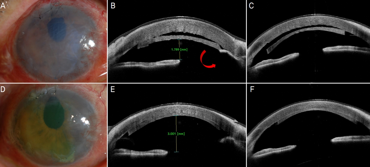
Slit-lamp and anterior segment optical coherence tomography (OCT) pictures of case. (A-C) Graft lenticle was detached from posterior stroma. Shallow anterior chamber and air migration to the posterior chamber (red arrow) were found after Descemet stripping automated endothelial keratoplasty (DSAEK). At postoperative 3 days, (D) cornea was maintained clear. (E, F) Deep anterior chamber and well attached graft lenticle was observed in anterior segment OCT.
수술 후 2개월 경과 관찰 결과 이식편의 분리, 급성 거부 반응, 각막미란 및 궤양 등의 합병증은 관찰되지 않았고 세극등현미경에서 중심부 각막은 투명해 보였다. 환자의 주관적 시력 호전은 있었으나 수술 후 회복 기간이 충분치 않아 장기간의 시력 예후는 경과 관찰이 필요할 것으로 보이나, 이전의 망막박리로 인한 시세포의 손상으로 인해 시력 회복에는 한계가 있을 것으로 생각된다.
고 찰
DSAEK은 수포각막병증, 푹스이영양증 등의 각막내피세포 부전 및 기능 감소가 있으며 각막상피 및 기질의 이상이 없을 시 선호되는 수술 방법으로 자리잡고 있다.3 PKP에 비해 술 후 시력 및 창상 회복이 빠르다는 장점이 있지만 합병증 또한 존재하는 수술이다.4,5 DSAEK의 가장 흔한 합병증은 이식편의 분리이다.6 특히 녹내장수술, 무홍채증, 홍채 변형, 무수정체, 유리체절제술을 시행한 안에서는 공기 방울을 전방에 잔류시키기 힘들기 때문에 이식편의 결합에 어려움이 따른다.7 본 증례 또한 유리체절제술을 시행하였고 홍채 변형이 있는 눈에서 DSAEK 시행 후 전방 내 공기가 유지되지 않아 이식편의 분리가 일어난 경우로, 강력한 산동제인 아트로핀을 이용하여 성공적인 재유착을 이뤄냈다는 점에서 의의를 찾을 수 있겠다.
본 증례와 같이 아트로핀을 이용하여 전방 내 공기 방울의 잔류를 유도하여 이식편을 재유착시킨 국내외 보고는 찾을 수 없었으나, Vaddavalli et al8의 연구에서는 각막 통기 절개(corneal venting incision) 및 술 후 높은 전방 내 안압 유지를 이식편의 결합을 유도하는 요소로 저술하고 있고, Koo9의 증례보고에서는 홍채 변형, 홍채 결손이 있는 환자에서 DSAEK 수술 중 공기 방울의 안정적인 전방으로의 주입을 위해 ‘sheet glide’를 사용하여 후방으로 공기의 이동을 막는 수술 기법을 소개하였다. 아트로핀은 DSAEK의 수술 후 합병증 중 하나인 동공 차단을 방지하는 역할을 위해 사용되기도 하지만, 본 증례에서는 수술 후 후방으로의 공기 이동이 관찰되어 이를 방지하기 위해 전방과 후방을 동역학적으로 하나의 공간으로 만들기 위한 방법으로 아트로핀을 사용하였다. 수술 후 1일째 전안부 빛간섭단층촬영에서 전방이 매우 얕고 동시에 이식편이 분리되어 있었기 때문에 실제로 공기가 전방에 잔류할 수 있는 공간은 더 좁은 상태였을 것으로 생각할 수 있겠다. 따라서 전방에 주입된 공기 방울에 가해지는 상대적인 압력이 커지게 되며 이때 홍채의 구조적 변형이 있는 부위로 공기의 흐름이 생긴 것으로 예상해 볼 수 있을 것이다. 그러므로 아트로핀 점안 후 전방이 깊어지고 공기 방울이 잔류할 수 있는 공간이 확보되면서 후방으로의 공기 이동이 관찰되지 않았고 이후 성공적으로 이식편이 유착된 것으로 생각된다.10 또한 본 증례처럼 홍채의 기능이 떨어져 있는 경우 가장 강력한 효과를 가진 아트로핀을 사용하는 것이 적절하였다고 보여진다. 따라서 전방에 구조적 기형이 있는 환자에게 동공차단 및 이식편 분리 등의 합병증을 줄일 수 있는 방법으로 아트로핀 점안을 고려해 볼 수 있겠다. 이때 아트로핀으로 인한 안압상승이 발생할 수 있으므로 각별히 주의 관찰이 필요할 것으로 보인다.11,12
한편, DSAEK 수술 시 이식편을 전방 내로 삽입하기 위해 글라이드를 이용하는 방법, 이식편을 접어 압력 최소화 포셉으로 삽입하는 방법, 인공수정체 삽입 카트리지에 이식편을 말아 넣어 삽입하는 방법 등이 보고되고 있다.13 Bradley and McCartney7의 연구에서 소개하고 있는 suture drag 기법은 홍채의 변형이 있거나 전방이 얕은 경우, 안압이 높은 경우에 이식편을 전방으로 삽입할 때 장점이 있으며, Sarnicola et al14의 연구에서는 suture pull-through 기법을 사용할 때 포셉을 사용하지 않기 때문에 삽입 과정에서 이식편의 손상이 적고, 전방을 유지하기 위한 장치 및 홍채 절개 등의 추가 시술이 필요하지 않아 술 후 합병증을 줄일 수 있다고 보고하였다. 본 증례에서는 suture pull-through 기법을 이용하여 이식편을 전방에 위치시킨 후 공기주입술을 시행하였는데, 이는 각막의 한쪽 끝이 봉합되어 있기 때문에 본 증례와 같이 이식편의 분리가 일어났을 때 그 위치가 어긋나는 것을 막아주는 역할 또한 기대할 수 있겠다. 이는 추후 공기재주입술 또는 이식편의 재유착을 위한 추가적 시술을 할 때에도 효과적인 접근을 가능케 할 것으로 생각된다.
결론적으로 홍채 변형과 같은 전방의 구조 이상이 있는 눈에서 DSAEK 시행 후 후방으로 공기 방울의 이동이 관찰될 경우 아트로핀과 같은 강력한 산동제를 이용하는 것이 도움이 될 수 있고, 이식편의 분리를 최소화하기 위해 suture pull-through 방법을 사용하는 것이 유용할 것으로 판단된다. 이때 아트로핀 점안으로 인한 안압상승 및 동공 차단, 전방각폐쇄 여부를 주의 깊게 관찰하여야 할 것이다.12 본 증례의 한계점으로는 한 가지 예이기 때문에 일반화에는 제한적이고, 경과 관찰 기간이 다소 짧으며 수술 전 시력이 매우 낮은 상태였기 때문에 시력 예후를 평가하는 데 어려움이 있었다는 점이다. 향후 본 증례와 유사한 전방의 구조적 변형이 있는 눈에서 DSAEK을 시행한 경우를 분석하여 이식편의 분리에 영향을 미치는 세부 인자에 관한 연구도 필요할 것으로 생각된다.
Notes
Conflicts of Interest
The authors have no conflicts to disclose.
References
Biography
양승안 / Seung Ahn Yang
양산부산대학교병원 안과학교실
Department of Ophthalmology, Pusan National University Yangsan Hospital