결막 광범위큰B세포림프종
Conjunctival Diffuse Large B-cell Lymphoma
Article information
Abstract
목적
눈꺼풀구석에 발생한 결막 광범위큰B세포림프종 1예를 보고하고자 한다.
증례요약
49세 남자 환자가 20여 일 전부터 발생한 오른눈 아래눈꺼풀구석의 충혈과 이물감을 주소로 내원하였다. 안구통증이나 전신적인 발열, 체중 감소는 없었으며, 동반된 만성 질환도 없었다. 세극등현미경검사에서 아래눈꺼풀구석 및 안구결막에 연어살색의 융기된 비교적 경계가 명확한 종양이 관찰되었다. 결막종양을 절제생검하였다. 육안검사에서 크기는 17×5×4 mm였다. 현미경검사에서 작고 성숙한 단핵림프구가 밀집된 구역과 분할된 핵과 명확한 핵소체를 보이는 큰 림프모세포와 같은 림프구가 밀집된 구역이 상존하였다. 면역조직화학염색에서 큰 세포 밀집구역은 CD20, Bcl-6 양성 소견과 CD3, CD5, CD23 음성을 보였고 상대적으로 높은 Ki-67 양성률을 보였다. 최종적으로 점막관련림프조직림프종에 동반된 광범위큰B세포림프종으로 진단받았다. 전신검사상 결막 이외의 다른 곳의 병변은 없었다. 환자는 절제생검 이후 항암화학요법을 받았으며 1년 뒤 외래 경과 관찰에서 재발은 없었다.
결론
결막림프종 환자에서 드물지만 광범위큰B세포림프종이 있을 수 있으므로 감별진단에 이를 염두에 두어야 한다.
Trans Abstract
Purpose
To report a case of diffuse large B-cell lymphoma of the conjunctival fornix.
Case summary
A 49-year-old man visited our clinic with redness and foreign body sensation in the right inferior conjunctival fornix that had begun 20 days previously. The patient had no pain, no fever, weight loss, and no past history of chronic disease. On slit lamp biomicroscopic examination, a large, salmon-colored, raised, well-defined, mass was detected in the inferior bulbar and fornix conjunctiva. We performed excision biopsy. The soft tissue lesion was 17 × 5 × 4 mm in size. Microscopic examination identified an area of small, mature mononuclear lymphocytes and an area of lymphocytes, such as large lymphoblasts with divided nuclei and conspicuous nucleoli. On immunohistochemical staining, the diffuse large B-cell lymphoma areas were positive for CD20 and Bcl-6, negative for CD3, CD5, and CD23, and the Ki-67 positive rate was relatively high. Finally, the patient was diagnosed with diffuse large B-cell lymphoma accompanying mucosal-associated lymphoid tissue lymphoma. There were no findings suggestive of metastasis invasion from other organs. The patient underwent immunochemotherapy after excisional biopsy. No recurrence has occurred over 1-year follow-up.
Conclusions
Although rare, diffuse large B-cell lymphoma should be considered in the differential diagnosis of conjunctival lymphomatous lesions.
림프종은 가장 흔한 악성결막종양 중의 하나다.1 한국인의 결막종양은 양성 종양이 가장 흔하지만, 악성 종양 중에서는 림프종이 가장 흔하고 90% 이상이 B림프구에서 기인한다.2,3 결막림프종 중에서는 점막관련림프조직림프종(mucosaassociated lymphoid tissue lymphoma)이 가장 흔하다.2,3 광범위큰B세포림프종(diffuse large B-cell lymphoma, DLBCL)은 비호지킨림프종(non-Hodgkin lymphoma)의 가장 흔한 형태로 눈부속기림프종(ocular adnexal lymphoma)의 5-15% 빈도로 발생하며 호발 위치는 눈확(orbit)이다.4,5 광범위큰B세포림프종은 빠르게 커지고 공격적 임상 경과를 보여 일반적으로 예후가 나쁘다.5,6 결막에 발생한 광범위큰B세포림프종은 매우 드물어 눈꺼풀판결막에서 발생한 몇몇 보고만 있다.7,8
아래눈꺼풀구석 및 안구결막에서 빠른 성장을 보이는 광범위큰B세포림프종은 국내에서는 아직 보고된 바 없어 이를 보고하고자 한다.
증례보고
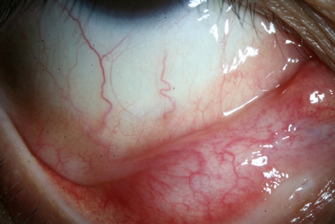
49세 남자 환자가 20일 전부터 발생한 오른눈의 충혈, 이물감으로 눈 아래쪽의 결막덩어리를 발견 후 본원에 내원하였다. 환자는 외상이나 전신감염은 없었으며 과거력상 특이 소견은 없었다. 초진 시 최대교정시력은 오른눈 0.8, 왼눈 1.0이었고, 안압은 정상이었다. 세극등현미경검사상 각막에 특이 소견은 없었으며, 오른눈의 아래결막구석에서 안구결막에 걸쳐 짙은 연어살색의 경계가 명확한 종양이 관찰되었다(Fig. 1). 종양은 내측눈구석에서 외측눈구석에 이르는 큰 크기의 혈관화된 종양이었다.

Anterior segment photograph of the everted right lower eyelid. Well-circumscribed, pink, vascular mass along inferior conjunctival fornix was visible.
국소마취 하에 결막 종양을 절제생검하였다. 육안검사상 연어살색을 띠는 타원형의 연조직으로 크기는 17×4×3 mm였다. 현미경검사상 결막 밑에 많은 림프구 침윤이 관찰되었는데 종양세포의 크기가 작고 핵소체가 잘 보이지 않은 세포가 밀집한 곳과 크기가 크고 중심모세포 또는 면역모세포와 같은 뚜렷한 핵소체를 보이는 곳이 섞여있었다. 면역화학염색상 작은 세포가 있는 곳에서는 CD20, Bcl-6, Bcl-2에 양성이었고 CD3, CD5, CD10, CD23, cyclin D1에는 음성이었으며 cytokeratin에는 양성이었으며 Ki-67 양성률이 3%로 낮았다. 반면, 큰 세포가 있는 곳에서는 CD20, Bcl-6에 양성이었고, CD3, CD5, CD10, CD23, cyclin D1, cytokeratin에는 음성이었으며 Ki-67은 25%로 상대적으로 높은 소견을 보였다. 악성림프종 중 점막관련림프조직림프종에 동반된 광범위큰B세포림프종(DLBCL)으로 확진되었다(Fig. 2).
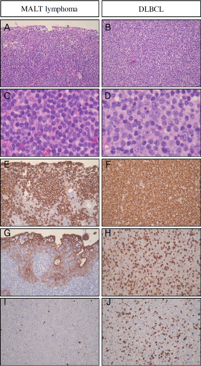
Histologic findings of diffuse large B-cell lymphoma accompanying mucosa-associated lymphoid tissue (MALT) lymphoma. (A, C) In the MALT lymphoma area, tumor cells infiltrate overlying epithelium and form lymphoepithelial lesion (A), and the tumor cells are small, and the nucleoli are inconspicuous (C). (B, D) The diffuse large B-cell lymphoma (DLBCL) area is composed of transformed centroblast-like or immunoblast-like large cells with conspicuous nucleoli. (E, F) The tumor cells in both MALT lymphoma and DLBCL area are positive for CD20. (G) Immunostaining for cytokeratin highlights lymphoepithelial lesions. (H) The tumor cells in the DLBCL area are negative for CD3. (I, J) The MALT lymphoma area show a low number of Ki67-positive cells (I), but the DLBCL area shows more number of Ki-67 positive cells. (A, B: Hematoxylin and Eosin [H&E] stain, original magnification ×400; C, D: H&E stain, digital magnification of ×400; E-J: Immunostain, original magnification ×400).
타원에서 시행한 전신검사상 전이 소견은 없었으며 추가로 rituximab, cyclophosphamide, doxorubicin, vincristine, prednisolone (R-CHOP) 항암화학요법을 받았다. 환자는 생검 이후 특별한 불편감을 호소하지 않았으며 1년 6개월 뒤 경과 관찰까지 재발의 소견은 없었다(Fig. 3).
Photograph the everted right lower eyelid after 1 year of immunochemotherapy. There was no tumor recurrence.
본 연구는 전북대학교병원 생명의학연구윤리심의위원회로부터 심의 면제를 받았다(승인 번호: CUH 2022-07-034).
고 찰
원발광범위큰B세포림프종(primary DLBCL)은 흔하지 않은 종양으로 최근 국내 단일 3차병원에서 보고한 눈과 눈부속기 원발림프종의 4.6%에서만이 발생하였다.6 결막에서의 발생은 매우 드물어 이제까지 3개 증례보고만 있다. Robinson et al7은 40세 남자에서 위눈꺼풀결막의 무통성의 큰 돌출된 덩어리로 보고하였고, Kim and Khwarg8은 55세 남자에서 아래눈꺼풀결막의 평평하고 매끈한 덩어리로 보고하였으며 Huerva et al9은 80세 여자에서 아래눈꺼풀결막에 중심부가 궤양처럼 패인 원형의 덩어리로 보고하였다. 본 증례에서는 빠르게 성장하는 아래눈꺼풀구석에서 발생한 돌출된 연어살색의 덩어리였다.
결막림프종은 비교적 예후가 양호한 것을 알려져 있지만 예후를 결정하는 가장 중요한 인자는 조직학적 아형으로 저등급(low-grade)인 점막관련림프조직림프종이나 소포림프종(follicular lymphoma)일 경우에 예후가 좋다.
광범위큰B세포림프종은 고등급(high-grade)으로 림프종 중 공격적인 경과를 보여 예후가 나쁜 아형이다. 광범위큰 B세포림프종은 발생 위치에 따라 예후가 달라지는데 Ahmed et al10은 유리체-망막이나 섬모체에 발생한 림프종의 경우 전체 생존 기간이 가장 짧았고, 눈확이나 눈꺼풀에 발생한 경우는 중간 예후를 보이고, 결막이나 눈물샘에서 발생한 경우가 가장 좋은 예후를 보인다고 하였다. Han et al6은 유리체 망막에 생긴 광범위큰B세포림프종의 경우 재발률이 높고 다른 전신으로 파급된 경우가 많아 생존율이 낮다고 하였다. 본 증례에서는 결막에만 발생하고 다른 전신 림프종은 없는 Ann Arbor stage 1E였다.
국소적으로 발생한 림프절 외 B세포림프종이거나 stage 1E이고 저등급인 눈부속기림프종의 우선적 치료는 모두 국소방사선 치료이다. 방사선 치료의 효과는 좋은 것으로 알려져 있으나 안구건조증, 각막궤양, 백내장, 방사선망막증과 같은 부작용의 우려가 있다. 수술적 절제로 치료한 경우도 보고되었으나 눈부속기 림프종의 경우 종양의 미세침윤 및 주변조직으로의 전파 우려로 단독 치료로는 권고되지 않는다.9 원발광범위큰B세포림프종을 마이토마이신C 0.04% 안약 점안으로 덩어리의 감소를 보였다는 보고도 있지만 R-CHOP 면역항암화학요법이 국소적인 눈 부작용을 줄이고 생존율을 높일 수 있다.7 본 증례에서도 40대의 젊은 환자여서 방사선 치료의 부작용이 우려되어 시행하지 않고 절제생검 후 R-CHOP 면역항암화학요법을 시행받았고 1년 6개월 이상 외래 경과에서 재발은 없었다.
결막에 발생한 광범위큰B세포림프종의 보고는 한국인에서는 아직 보고된 바 없다. 따라서 결막에서 발생하는 연어살색의 림프종에서 드물게 발생하지만 이에 대한 감별이 필요하다.
Notes
Conflicts of Interest
The authors have no conflicts to disclose.
References
Biography
김영은 / Young-Eun Kim
전북대학교 의과대학 안과학교실
Department of Ophthalmology, Jeonbuk National University Medical School
