 |
 |
| J Korean Ophthalmol Soc > Volume 63(4); 2022 > Article |
|
ĻĄŁļ¼Ėņ┤łļĪØ
ļ¬®ņĀü
ļ¦Øļ¦ēņāēņåīļ│Ćņä▒ņ£╝ļĪ£ ņ¦äļŗ©ļÉ£ ĻĄŁļé┤ ĒÖśņ×ÉņØś ņ×äņāüņĀü ĒŖ╣ņä▒ņØä ņĢīņĢäļ│┤Ļ│Āņ×É ĒĢ£ļŗż.
ļīĆņāüĻ│╝ ļ░®ļ▓Ģ
2014ļģä 1ņøöļČĆĒä░ 2019ļģä 12ņøö ņé¼ņØ┤ņŚÉ ļ¦Øļ¦ēņāēņåīļ│Ćņä▒ņ£╝ļĪ£ ņĄ£ņ┤ł ņ¦äļŗ©ļ░øņØĆ ĒÖśņ×Éļź╝ ļīĆņāüņ£╝ļĪ£ ĒøäĒ¢źņĀü ņØśļ¼┤ĻĖ░ļĪØ ļČäņäØņØä ņŗ£Ē¢ēĒĢśņśĆļŗż. ĒÖśņ×ÉņØś ļéśņØ┤, ņä▒ļ│ä, ņŻ╝ļÉ£ ņåīĻ▓¼ ļ░Å ņĄ£ļīĆĻĄÉņĀĢņŗ£ļĀźņØä ĒÖĢņØĖĒĢśņśĆļŗż. ņČöĻ░ĆļĪ£ ĒøäļéŁĒĢśļ░▒ļé┤ņן ņŚ¼ļČĆ, ļ╣øĻ░äņäŁļŗ©ņĖĄņ┤¼ņśüņāüņØś ņØ┤ņāü ņåīĻ▓¼, ņŗ£ņĢ╝Ļ▓Ćņé¼, ļłłņĀäņ£äļÅäĻ▓Ćņé¼ņØś Ļ▓░Ļ│╝ņŚÉ ļīĆĒĢ£ ļČäņäØņØä ņŗ£Ē¢ēĒĢśņśĆļŗż.
Ļ▓░Ļ│╝
ņ┤Ø 246ļ¬ģ 492ņĢłņØä ļīĆņāüņ£╝ļĪ£ Ļ▓░Ļ│╝ļź╝ ļČäņäØĒĢśņśĆļŗż. ĒÅēĻĘĀ ļéśņØ┤ļŖö 48.0 ┬▒ 16.0ņäĖņśĆņ£╝ļ®░, ņŻ╝ļÉ£ ņŻ╝ĒśĖņåīļŖö ņŗ£ļĀźņĀĆĒĢśņÖĆ ņĢ╝ļ¦╣ņ”ØņØ┤ņŚłļŗż. ĒÅēĻĘĀ logMAR ņĄ£ļīĆĻĄÉņĀĢņŗ£ļĀźņØĆ 0.31 ┬▒ 0.50ņØ┤ņŚłņ£╝ļ®░ 368ņĢł(74.8%)ņŚÉņä£ decimal ņŗ£ļĀźņØ┤ 0.5 ņØ┤ņāüņØ┤ņŚłļŗż. ņŗ£ņĢ╝Ļ▓Ćņé¼ļź╝ ņŗ£Ē¢ēĒĢ£ 462ņĢł ņżæ 328ņĢł(71.0%)ņŚÉņä£ ņżæņŗ¼ 10┬░ ņØ┤ļé┤ņØś ņŗ£ņĢ╝ņØ┤ņāüņØ┤ ļéśĒāĆļé¼ņ£╝ļ®░, ļłłņĀäņ£äļÅäĻ▓Ćņé¼ļź╝ ņŗ£Ē¢ēĒĢ£ 242ņĢłņŚÉņä£ ĒÅēĻĘĀ ņĢäļŹ┤ļ╣äļŖö 1.28 ┬▒ 0.28ņØ┤ņŚłļŗż. ļ╣øĻ░äņäŁļŗ©ņĖĄņ┤¼ņśüņŚÉņä£ ņ£Āļ”¼ņ▓┤ĒÖ®ļ░śĻ▓¼ņØĖ/ļ¦Øļ¦ēņĢ×ļ¦ēņØ┤ 135ņĢł(27.4%), ļéŁĒżĒÖ®ļ░śļČĆņóģņØ┤ 48ņĢł(9.8%), ĒÖ®ļ░śņØś ņ¢ćņĢäņ¦ÉņØ┤ 112ņĢł(22.8%)ņŚÉņä£ Ļ┤Ćņ░░ļÉśņŚłļŗż. ņżæņŗ¼ņÖĆ ĒāĆņøÉņ▓┤ĻĄ¼ņŚŁ ņāüĒā£ņØś Ļ▓ĮņÜ░ ņś©ņĀäĒĢ£ ņāüĒā£Ļ░Ć 222ņĢł(45.1%), ņåÉņāüļÉ£ ņāüĒā£Ļ░Ć 220ņĢł(44.7%), ņŗ¼ĒĢ£ ņåÉņāüņ£╝ļĪ£ Ļ┤Ćņ░░ļÉśņ¦Ć ņĢŖļŖö ņāüĒā£Ļ░Ć 50ņĢł(10.2%)ņØ┤ņŚłļŗż.
ABSTRACT
Purpose
To evaluate the clinical characteristics of Korean patients diagnosed with retinitis pigmentosa.
Methods
We retrospectively reviewed the medical records of patients diagnosed with retinitis pigmentosa from January 2014 to December 2019. We evaluated age, gender, the chief complaints, posterior subcapsular cataract status, abnormalities on optical coherence tomography, visual field test results, and electrooculograms.
Results
A total of 492 eyes of 246 patients were included. The mean patient age was 48.0 ┬▒ 16.0 years and the chief complaints were decreased vision and night blindness. The mean logarithm of the minimal angle of resolution (logMAR) bestŌĆÉcorrected visual acuity (BCVA) was 0.31 ┬▒ 0.50. The BCVA was 0.5 or better in 368 eyes (74.8%). A total of 328 (71.0%) of 462 eyes that underwent visual field testing exhibited visual field defects within 10┬║. The mean Arden ratio was 1.28 ┬▒ 0.28 for the 242 eyes that underwent electroŌĆÉoculography. Optical coherence tomography revealed vitreomacular traction/an epiretinal membrane, cystoid macular edema, and retinal thinning in 135 (27.4%), 48 (9.8%), and 112 (22.8%) eyes, respectively. The ellipsoid zone was intact in 222 eyes (45.1%), disrupted in 220 (44.7%), and absent in 50 (10.2%).
ļ¦Øļ¦ēņāēņåīļ│Ćņä▒ņØĆ ņĀÉņ¦äņĀüņØĖ ļ¦Øļ¦ēņāēņåīņāüĒö╝ņÖĆ ņŗ£ņäĖĒżņĖĄņØś ļ│Ćņä▒ņØä ĒŖ╣ņ¦Ģņ£╝ļĪ£ ĒĢśļŖö ņ£ĀņĀäņä▒ ļ¦Øļ¦ēņ¦łĒÖśņØ┤ļŗż[1]. ĻĄŁļé┤ ļ░£ņāØļźĀņØĆ 100,000ļ¬ģļŗ╣ ņĢĮ 1.63ŌĆÉ1.64ļ¬ģ ņĀĢļÅäļĪ£ ņĢīļĀżņĀĖ ņ׳ņ£╝ļ®░, 20ŌĆÉ24ņäĖ ļ░Å 65ŌĆÉ69ņäĖņŚÉ ļ░£ņāØļźĀņØ┤ ņāüļīĆņĀüņ£╝ļĪ£ ļåÆĻ▓ī ļéśĒāĆļé¼ļŗż[2]. Na et al. [3]ņØś ņŚ░ĻĄ¼ņŚÉ ļö░ļź┤ļ®┤ 2011ŌĆÉ2014ļģäņØś ņŚ░ĻĄ¼ ĻĖ░Ļ░ä ļÅÖņĢł ĻĄŁļé┤ ņ£Āļ│æļźĀņØĆ 100,000ļ¬ģļŗ╣ 11.09ļ¬ģņ£╝ļĪ£ ļéśĒāĆļé¼ļŖöļŹ░, 40ņäĖ ņØ┤ņāüņŚÉņä£ļŖö 100,000ļ¬ģļŗ╣ 16.16ļ¬ģņ£╝ļĪ£ ņ£Āļ│æļźĀņØ┤ ņāüļīĆņĀüņ£╝ļĪ£ ļåÆņØĆ Ļ▓ĮĒ¢źņØä ļ│┤ņśĆļŗż. Ļ│╝Ļ▒░ ĒĢ┤ņÖĖ ļ│┤Ļ│ĀņŚÉņä£ļŖö 3,000ŌĆÉ5,000ļ¬ģļŗ╣ 1ļ¬ģ ņĀĢļÅäņØś ņ£Āļ│æļźĀņØä ļ│┤ņØ┤ļŖö Ļ▓āņ£╝ļĪ£ ņĪ░ņé¼ļÉśņŚłļŗż[4]. ļ¦Øļ¦ēņāēņåī ļ│Ćņä▒ņØ┤ ņ¦äĒ¢ēļÉśļŖö Ļ▓ĮņÜ░ ņĢ╝ļ¦╣ņ”Ø, ņŗ£ņĢ╝ņןņĢĀ ļ░Å ņŗ£ļĀźņĀĆĒĢśļź╝ ņ£Āļ░£ĒĢśņŚ¼ ņŗ¼ĒĢ£ Ļ▓ĮņÜ░ ņŗżļ¬ģņŚÉ ņØ┤ļź╝ ņłś ņ׳ņ£╝ļ®░[5]. Ēśäņ×¼Ļ╣īņ¦Ć ĻĘĖ ņ¦äĒ¢ēņØä ļŖ”ņČöĻ▒░ļéś ņ¦łĒÖśņØä ņÖäņ╣śņŗ£Ēé¼ ņłś ņ׳ļŖö ĒÖĢļ”ĮļÉ£ ļ░®ļ▓ĢņØ┤ ņŚåļŖö ņāüĒā£ņØ┤ļŗż.
ņĄ£ĻĘ╝ ņ£ĀņĀäņ×É ļČäņäØ ļ░®ņŗØņØś ļ░£ļŗ¼ņŚÉ ļö░ļØ╝ ļ¦Øļ¦ēņāēņåīļ│Ćņä▒Ļ│╝ Ļ┤ĆļĀ©ļÉ£ ļ│┤ļŗż ļ¦ÄņØĆ ņ£ĀņĀä ņØ┤ņāüļōżņØ┤ ļ░ØĒśĆņ¦ĆĻ│Ā ņ׳ņ£╝ļ®░[5], ļŗżņ¢æĒĢ£ ļ░®ņŗØņØś ņ£ĀņĀäņ×É ņ╣śļŻīņŚÉ ļīĆĒĢ£ ņŗżĒŚśņĀü ņŗ£ļÅäļōżņØ┤ ņØ┤ļŻ©ņ¢┤ņ¦ĆĻ│Ā ņ׳ļŗż[1]. ļŹö ļéśņĢäĻ░Ć ņ£ĀņĀäņä▒ ņ¦łĒÖśņŚÉ ļīĆĒĢ£ ņāüņŚģņĀüņØĖ ņ£ĀņĀäņ×É ļČäņäØņØ┤ ļ│┤ļŗż ļäÉļ”¼ ņŗ£Ē¢ēļÉśļŖö ņČöņäĖ[6]ņŚÉ ļö░ļØ╝ ļ¦Øļ¦ēņāēņåīļ│Ćņä▒ņØ┤ ņØśņŗ¼ļÉśļŖö ĒÖśņ×ÉņŚÉ ļīĆĒĢ£ ļ│┤ļŗż ņĀüĻĘ╣ņĀüņØĖ ņ£ĀņĀäņ×ÉĻ▓Ćņé¼ņÖĆ ņ£ĀņĀä ņāüļŗ┤(genetic counseling)ņØ┤ ĒÖ£ņä▒ĒÖöļÉĀ Ļ▓āņ£╝ļĪ£ ĻĖ░ļīĆļÉśĻ│Ā ņ׳ļŗż.
ņ¢┤ļ¢ż ņ¦łĒÖśņ£╝ļĪ£ ņ¦äļŗ©ļÉ£ ĒÖśņ×ÉņÖĆ ņāüļŗ┤ņØä ņŗ£Ē¢ēĒĢśļŖö ļŹ░ņŚÉ ņ׳ņ¢┤ņä£ ņāüĻĖ░ ņ¦łĒÖśņØä Ļ░Ćņ¦ä ņŚ¼ļ¤¼ ĒÖśņ×ÉļōżņØś ņØ╝ļ░śņĀüņØĖ ĒŖ╣ņä▒ņŚÉ ļīĆĒĢ£ ņĀĢļ│┤ļŖö ņ£ĀņÜ®ĒĢśĻ▓ī ņØ┤ņÜ®ļÉĀ ņłś ņ׳ļŗż. ļ│Ė ņŚ░ĻĄ¼ņŚÉņä£ļŖö 246ļ¬ģņØ┤ļØ╝ļŖö ļ╣äĻĄÉņĀü ļ¦ÄņØĆ ĻĄŁļé┤ ĒÖśņ×ÉļōżņØä ļīĆņāüņ£╝ļĪ£ ĻĘĖ ņ×äņāüņĀü ĒŖ╣ņä▒Ļ│╝ ļ╣øĻ░äņäŁļŗ©ņĖĄņ┤¼ņśü ņāü ĒŖ╣ņä▒ņØä ļČäņäØĒĢśĻ│Ā ņØ┤ļź╝ ļ│┤Ļ│ĀĒĢśĻ│Āņ×É ĒĢ£ļŗż.
ļ│Ė ĒøäĒ¢źņĀü ņŚ░ĻĄ¼ļŖö ļŗ©ņØ╝ ĻĖ░Ļ┤ĆņŚÉņä£ ĒŚ¼ņŗ▒ĒéżņäĀņ¢ĖņŚÉ ņ×ģĻ░üĒĢśņŚ¼ ņŗ£Ē¢ēļÉśņŚłņ£╝ļ®░, Institutional Review Board (IRB) ņŖ╣ņØĖņØä ĒÜŹļōØĒĢśņśĆļŗż(IRB No. 2020-05-001). 2014ļģä 1ņøöļČĆĒä░ 2019ļģä 12ņøöĻ╣īņ¦Ć ņĀäļ¼Ė ļ│æņøÉņØĖ Ļ╣ĆņĢłĻ│╝ļ│æņøÉņØä ļ░®ļ¼ĖĒĢśņŚ¼ ļ¦Øļ¦ēņāēņåīļ│Ćņä▒ņ£╝ļĪ£ ņĄ£ņ┤łļĪ£ ņ¦äļŗ©ļ░øņØĆ ĒÖśņ×Éļź╝ ļīĆņāüņ£╝ļĪ£ ņØśļ¼┤ĻĖ░ļĪØņØä ļČäņäØĒĢśņśĆļŗż. ņØ┤ņĀäņŚÉ ĒāĆ ļ│æņøÉņŚÉņä£ ļ¦Øļ¦ēņāēņåīļ│Ćņä▒ņØä ņ¦äļŗ©ļ░øņØĆ Ēøä Ļ▓ĮĻ│╝ Ļ┤Ćņ░░ĒĢśļŗż ņāüĒā£ ĒÅēĻ░Ćļź╝ ņ£äĒĢ┤ ļ│ĖņøÉņŚÉ ļé┤ņøÉĒĢ£ Ļ▓ĮņÜ░ļŖö ņŚ░ĻĄ¼ņŚÉņä£ ņĀ£ņÖĖĒĢśņśĆļŗż.
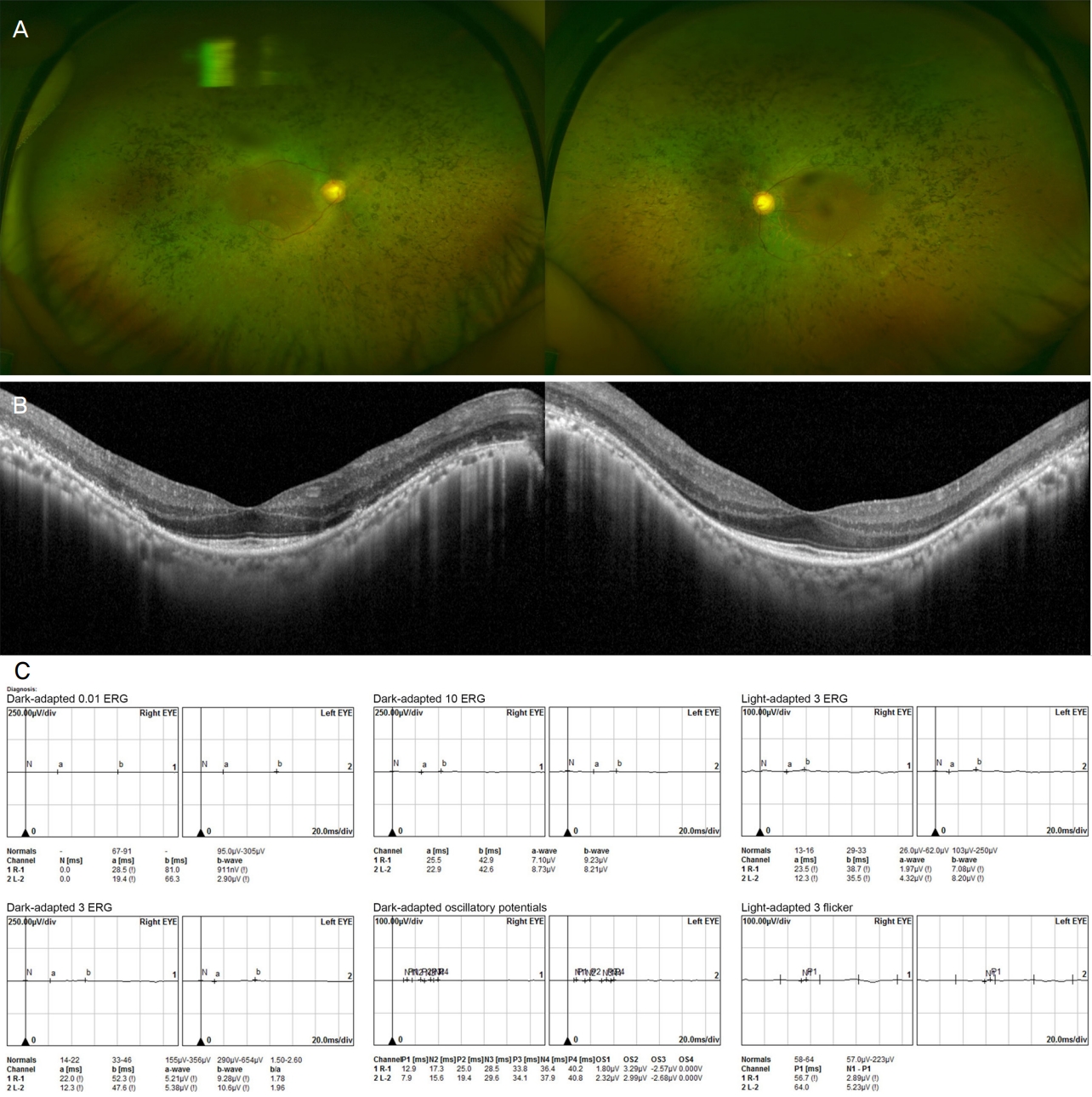
ņĢłņĀĆĻ▓Ćņé¼ ļ░Å ņĢłņĀĆņ┤¼ņśü Ļ▓░Ļ│╝, ļ¦Øļ¦ēņĀäņ£äļÅäĻ▓Ćņé¼(electroretinogram)ļź╝ ņØ┤ņÜ®ĒĢśņŚ¼ ņ¦łĒÖśņØä ņ¦äļŗ©ĒĢśņśĆņ£╝ļ®░, ļ╣øĻ░äņäŁļŗ©ņĖĄņ┤¼ņśü(optical coherence tomography) Ļ▓░Ļ│╝ļź╝ ņČöĻ░ĆņĀüņ£╝ļĪ£ ņ░ĖĻ│ĀĒĢśņśĆļŗż. ņĢłņĀĆĻ▓Ćņé¼ņŚÉņä£ ņŻ╝ļ│ĆļČĆ ļ¦Øļ¦ēņāēņåīņāüĒö╝ņØś Ēāłņāēņåīļéś ņ£äņČĢ, ļ╝łņĪ░Ļ░ü ļ¬©ņ¢æ ņāēņåīņ╣©ņ░®(bonyŌĆÉspicule pigmentation)ņØ┤ Ļ┤Ćņ░░ļÉśļ®░, ļ¦Øļ¦ēņĀäņ£äļÅäĻ▓Ćņé¼(electroretinogram)ņŚÉņä£ ļ¦ēļīĆņäĖĒż ļ░śņØæ ļśÉļŖö ļ¦ēļīĆņäĖĒżņÖĆ ņøÉļ┐öņäĖĒż ļ░śņØæņØś Ļ░ÉņåīĻ░Ć ļéśĒāĆļéśļ®┤ņä£ Ļ│╝Ļ▒░ ņŚ╝ņ”Ø, ņÖĖņāü ļśÉļŖö ņĢĮļ¼╝ ļō▒ņØś ņøÉņØĖņŚÉ ņØśĒĢ£ ņØ┤ņ░©ņĀüņØĖ ļ¦Øļ¦ēņØś ļ│ĆĒÖöļź╝ ļ░░ņĀ£ĒĢĀ ņłś ņ׳ļŖö Ļ▓ĮņÜ░ ļ¦Øļ¦ēņāēņåīļ│Ćņä▒ņ£╝ļĪ£ ņ¦äļŗ©ĒĢśņśĆļŗż(Fig. 1). ņČöĻ░ĆņĀüņ£╝ļĪ£ ļ╣øĻ░äņäŁļŗ©ņĖĄņ┤¼ņśüņŚÉņä£ ņØ┤ĒÖśļÉ£ ļČĆņ£äņØś ĒāĆņøÉņ▓┤ĻĄ¼ņŚŁ ņåÉņāüņØ┤ Ļ┤Ćņ░░ļÉśļŖö Ļ▓ĮņÜ░ ņ¦äļŗ©ņŚÉ ņ░ĖĻ│ĀĒĢśņśĆļŗż.
ļ¬©ļōĀ ĒÖśņ×ÉļŖö ņ¦äļŗ© ļŗ╣ņŗ£ Ēśäņä▒ĻĄ┤ņĀłĻ▓Ćņé¼, ņĄ£ļīĆĻĄÉņĀĢņŗ£ļĀź ņĖĪņĀĢ, ņäĖĻĘ╣ļō▒Ēśäļ»ĖĻ▓ĮĻ▓Ćņé¼, ņĢłņĀĆĻ▓Ćņé¼, ņĢłņĀĆņ┤¼ņśü, ļ╣øĻ░äņäŁļŗ©ņĖĄņ┤¼ņśü ļ░Å ļ¦Øļ¦ēņĀäņ£äļÅäĻ▓Ćņé¼ļź╝ ņŗ£Ē¢ēļ░øņĢśļŗż. ņČöĻ░ĆņĀüņ£╝ļĪ£ ņØśņé¼Ļ░Ć ļ¦Øļ¦ēņāēņåīņāüĒö╝ ĻĖ░ļŖźņŚÉ ļīĆĒĢśņŚ¼ ĒÖĢņØĖĒĢśĻ│Āņ×É ĒĢ£ Ļ▓ĮņÜ░ ļłłņĀäņ£äļÅäĻ▓Ćņé¼(electrooculogram)ļź╝ ņŗ£Ē¢ēļ░øņĢśņ£╝ļ®░, ĒÖśņ×ÉņØś ņŗ£ņĢ╝ ņØ┤ņāüņØä ĒÖĢņØĖĒĢśĻ│Āņ×É ĒĢ£ Ļ▓ĮņÜ░ ņŗ£ņĢ╝Ļ▓Ćņé¼ļź╝ ņŗ£Ē¢ēļ░øņĢśļŗż. ņĢłņĀĆņ×ÉĻ░ĆĒśĢĻ┤æ(fundus autofluorescence)ņØś Ļ▓ĮņÜ░ ņåīņłśņØś ĒÖśņ×ÉņŚÉņä£ļ¦ī ņŗ£Ē¢ēļÉśņŚłņ£╝ļ®░, Ļ▓░Ļ│╝ ļČäņäØņŚÉ ĒżĒĢ©ĒĢśņ¦Ć ņĢŖņĢśļŗż. Ļ░ĆņĪ▒ļĀźņØś Ļ▓ĮņÜ░ ņØśņé¼ņØś ĒīÉļŗ©ņŚÉ ļö░ļØ╝ ņäĀĒāØņĀüņ£╝ļĪ£ ĒÖĢņØĖĒĢśņśĆļŖöļŹ░, ĒÖśņ×ÉņØś ņ¦äņłĀņŚÉ ņØśĻ▒░ĒĢśņŚ¼ ņ£ĀņĀä ņ¢æņŗØņØä ĒÅēĻ░ĆĒĢśņśĆļŗż.
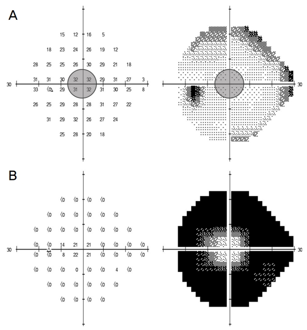
ļ╣øĻ░äņäŁļŗ©ņĖĄņ┤¼ņśüņŚÉļŖö ņä£ļĪ£ ļŗżļźĖ ņäĖ ĻĖ░ĻĖ░(Spectralis HRAOCT ┬«, Heidelberg Engineering, Heidelberg, Germany; RS 3000┬«, Nidek Co., Ltd., Tokyo, Japan; Spectral OCT┬«, Ophthalmic Technologies Inc., Toronto, Canada)Ļ░Ć ņØ┤ņÜ®ļÉśņŚłņ£╝ļ®░, ņŗ£ņĢ╝Ļ▓Ćņé¼ņØś Ļ▓ĮņÜ░ ĒŚśĒöäļ”¼(Humphrey) ļ░®ņŗØĻ│╝ Ļ│©ļō£ļ¦ī(Goldmann) ļ░®ņŗØņØ┤ Ēś╝ņÜ®ļÉśņŚłļŗż. ĒŚśĒöäļ”¼ ļ░®ņŗØņ£╝ļĪ£ Ļ▓Ćņé¼ĒĢ£ Ļ▓░Ļ│╝ņŚÉņä£ mean deviation (MD), macular sensitivity (MS)ļź╝ ņĖĪņĀĢĒĢśņśĆņ£╝ļ®░ MSļŖö ņżæņŗ¼ 4ņĀÉņØś sensitivityņØś ĒÅēĻĘĀĻ░Æņ£╝ļĪ£ ĻĄ¼ĒĢśņśĆļŗż(Fig. 2). ņŗ£ļĀźņØĆ decimal ņŗ£ļĀźĒæ£ļź╝ ņØ┤ņÜ®ĒĢśņŚ¼ ņĖĪņĀĢĒĢ£ Ēøä logarithm of minimal angle of resolution (logMAR) Ļ░Æņ£╝ļĪ£ ļ│ĆĒÖśĒĢśņśĆļŗż. ņĢłņĀäņłśņ¦ĆņÖĆ ņĢłņĀäņłśļÅÖņŗ£ļĀźņØś Ļ▓ĮņÜ░ Holladay7ņØś ņĀ£ņĢłņŚÉ ļö░ļØ╝ Ļ░üĻ░ü logMAR Ļ░Æ 2ņÖĆ 3ņ£╝ļĪ£ ļ│ĆĒÖśĒĢśņśĆļŗż. Ļ┤æĻ░üņ£Ā(light perception) ņŗ£ļĀźņØś Ļ▓ĮņÜ░ ņłśņ╣śĒÖöĒĢśĻĖ░ ņ¢┤ļĀżņÜ┤ ņŗ£ļĀźņ£╝ļĪ£ ņāØĻ░üļÉśĻ│Ā ņ׳ņ£╝ļéś[7], ļ│Ė ņŚ░ĻĄ¼ņŚÉņä£ļŖö ĒÅēĻĘĀ ņŗ£ļĀź ļČäņäØņØä ņ£äĒĢ┤ ņ×äņØśļĪ£ Ļ┤æĻ░üņ£Ā ņŗ£ļĀźņØä ņĢłņĀäņłśļÅÖĻ│╝ Ļ░ÖņØĆ logMAR Ļ░Æ 3ņ£╝ļĪ£ ļ│ĆĒÖśĒĢśņśĆļŗż.
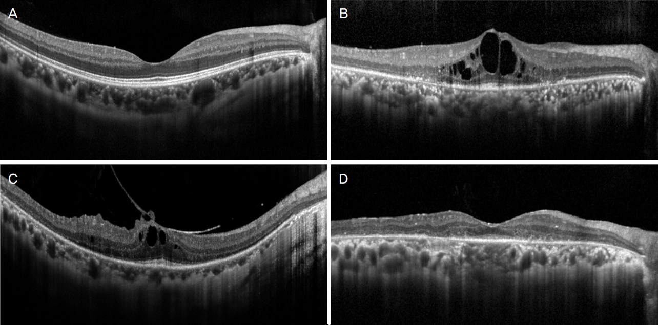
ņØśļ¼┤ ĻĖ░ļĪØ ļ░Å Ļ▓Ćņé¼ ĻĖ░ļĪØņØä ļČäņäØĒĢśņŚ¼ ļŗżņØīĻ│╝ Ļ░ÖņØĆ ĒĢŁļ¬®ņŚÉ ļīĆĒĢ£ ņĀĢļ│┤ļź╝ ņłśņ¦æĒĢśņśĆļŗż: ļéśņØ┤, ņä▒ļ│ä, ļŗ╣ļć©, Ļ│ĀĒśłņĢĢ, ĻĖ░ĒāĆ ņĀä ņŗĀņ¦łĒÖś, ļé£ņ▓Ł ņŚ¼ļČĆ, ļŗ©ņĖĪņä▒ ņŚ¼ļČĆ, ĻĄ┤ņĀł ņØ┤ņāü, ņĄ£ļīĆĻĄÉņĀĢņŗ£ļĀź, ĒøäļéŁĒĢśļ░▒ļé┤ņן(posterior subcapsular cataract) ņŚ¼ļČĆ, ļ╣øĻ░äņäŁ ļŗ©ņĖĄņ┤¼ņśüņāü ņØ┤ņāü ņåīĻ▓¼, ļéŁĒżĒÖ®ļ░śļČĆņóģ(cystoid macular edema) ļ░Å ĻĖ░ĒāĆ ĒÖ®ļ░śļČĆņØś ņØ┤ņāü ņŚ¼ļČĆ, ļ¦Øļ¦ēņĀäņ£äļÅäĻ▓Ćņé¼ ņżæ ņĢöņł£ņØæ(darkŌĆÉadapted) 3 Ļ▓Ćņé¼ņŚÉņä£ ņĖĪņĀĢļÉ£ aŌĆÉwaveņÖĆ bŌĆÉwaveņØś amplitude, ļłłņĀäņ£äļÅäĻ▓Ćņé¼ņāü ņĢäļŹ┤ļ╣ä(Arden ratio), ņŗ£ņĢ╝Ļ▓Ćņé¼ņāü ņØ┤ņāü ņåīĻ▓¼ ņŚ¼ļČĆ. ļ╣øĻ░äņäŁļŗ©ņĖĄņ┤¼ņśü ņØ┤ļ»Ėņ¦ĆņāüņØś ņØ┤ņāü ņåīĻ▓¼ņØś Ļ▓ĮņÜ░ Lupo et al [8]ņØś ņŚ░ĻĄ¼ņŚÉņä£ņÖĆ Ļ░ÖņØ┤ 4Ļ░£ groupņ£╝ļĪ£ ĻĄ¼ļČäĒĢśņśĆļŗż: group 1=ĒÖ®ļ░śņØś ĒŖ╣ņØ┤ ņØ┤ņāü ņåīĻ▓¼ņØ┤ ņŚåļŖö Ļ▓ĮņÜ░, group 2=ļéŁĒżĒÖ®ļ░śļČĆņóģņØ┤ ņ׳ļŖö Ļ▓ĮņÜ░, group3=ņ£Āļ”¼ņ▓┤ĒÖ®ļ░śĻ▓¼ņØĖ/ļ¦Øļ¦ēņĢ×ļ¦ēņØ┤ ņ׳ļŖö Ļ▓ĮņÜ░, group 4=ĒÖ®ļ░śņØś ņ¢ćņĢäņ¦É(thinning)ņØ┤ ņ׳ļŖö Ļ▓ĮņÜ░(Fig. 3). ņČöĻ░ĆņĀüņ£╝ļĪ£ ļ╣øĻ░äņäŁļŗ©ņĖĄņ┤¼ņśü ņØ┤ļ»Ėņ¦Ćļź╝ ļČäņäØĒĢśņŚ¼ ņżæņŗ¼ņÖĆ ĒāĆņøÉņ▓┤ĻĄ¼ņŚŁņØś ņāüĒā£ļź╝ ņś©ņĀäĒĢ£ ņāüĒā£(intact), ņåÉņāüļÉ£ ņāüĒā£(disrupted), ņŗ¼ĒĢ£ ņåÉņāüņ£╝ļĪ£ Ļ┤Ćņ░░ļÉśņ¦Ć ņĢŖļŖö ņāüĒā£(absent)ļĪ£ ĻĄ¼ļČäĒĢśņśĆļŗż(Fig. 4).
ņĀäņ▓┤ ĒÖśņ×Éļź╝ ļéśņØ┤ņŚÉ ļö░ļØ╝ 30ņäĖ ļ»Ėļ¦īĻĄ░, 30-50ņäĖĻĄ░, 50ņäĖ ņØ┤ņāüĻĄ░ņØś ņäĖ ĻĄ░ņ£╝ļĪ£ ĻĄ¼ļČäĒĢśņśĆļŗż. ņäĖ ĻĄ░ ņé¼ņØ┤ņŚÉ ņŗ£ļĀź, ņŗ£ņĢ╝ Ļ▓Ćņé¼ņāü MD ļ░Å MS, ļłłņĀäņ£äļÅäĻ▓Ćņé¼ņāü ņĢäļŹ┤ļ╣äļź╝ ņä£ļĪ£ ļ╣äĻĄÉĒĢś ņśĆņ£╝ļ®░, ņČöĻ░ĆņĀüņ£╝ļĪ£ ĒāĆņøÉņ▓┤ĻĄ¼ņŚŁņØś ņåÉņāü ņĀĢļÅä(ņś©ņĀäĒĢ£ ņāüĒā£ vs. ņåÉņāüļÉ£ ņāüĒā£ vs. ņŗ¼ĒĢ£ ņåÉņāüņ£╝ļĪ£ Ļ┤Ćņ░░ļÉśņ¦Ć ņĢŖļŖö ņāüĒā£)ļź╝ ļ╣äĻĄÉĒĢśņśĆļŗż.
ĒåĄĻ│ä ļČäņäØņŚÉļŖö SPSS ĒöäļĪ£ĻĘĖļש(SPSS ver. 25.0 for Windows;IBM Corp., Armonk, NY, USA)ņØä ņØ┤ņÜ®ĒĢśņśĆļŗż. ņŗ£ļĀźņØĆ logMAR Ļ░ÆņØä ņØ┤ņÜ®ĒĢśņŚ¼ ļČäņäØĒĢśņśĆļŗż. ņä£ļĪ£ ļŗżļźĖ ņäĖ ĻĄ░ ņé¼ņØ┤ņØś ļ╣äĻĄÉņŚÉļŖö oneŌĆÉway analysis of variances with Tukey test Ēś╣ņØĆ chiŌĆÉsquare testļź╝ ņØ┤ņÜ®ĒĢśņśĆņ£╝ļ®░, 0.05 ļ»Ėļ¦īņØś pĻ░ÆņØä ĒåĄĻ│äņĀüņ£╝ļĪ£ ņ£ĀņØśĒĢ£ Ļ░Æņ£╝ļĪ£ ņĀĢņØśĒĢśņśĆļŗż.
ņŚ░ĻĄ¼ ĻĖ░Ļ░ä ļÅÖņĢł ņĀäņ▓┤ 246ļ¬ģ(492ņĢł)ņØś ĒÖśņ×ÉĻ░Ć ļ¦Øļ¦ēņāēņåīļ│Ćņä▒ņ£╝ļĪ£ ņ¦äļŗ©ļÉśņŚłļŗż(Table 1). ļé©ņä▒ 97ļ¬ģ, ņŚ¼ņä▒ 149ļ¬ģņØ┤ņŚłņ£╝ļ®░, ļŗ╣ļć©, Ļ│ĀĒśłņĢĢņØĆ Ļ░üĻ░ü 21ļ¬ģ, 38ļ¬ģņŚÉņä£ ņ¦äļŗ©ļÉśņŚłļŗż. ĒÅēĻĘĀ ļéśņØ┤ļŖö 48.0 ┬▒ 16.0ņäĖņśĆņ£╝ļ®░, ĒÖśņ×ÉņØś ņŻ╝ ņ”ØņāüņØĆ ņŗ£ļĀźņĀĆĒĢśĻ░Ć 53ļ¬ģ(21.6%), ņĢ╝ļ¦╣ņ”ØņØ┤ 64ļ¬ģ(26.0%), Ļ┤æņŗ£ņ”ØņØ┤ 1ļ¬ģ(0.4%), ņŗ£ņĢ╝ ņØ┤ņāüņØ┤ 10ļ¬ģ(4.1%), ņŗ£ļĀźņĀĆĒĢśņÖĆ ņĢ╝ļ¦╣ņ”ØņØä Ļ░ÖņØ┤ ĒśĖņåīĒĢ£ Ļ▓ĮņÜ░Ļ░Ć 65ļ¬ģ(26.4%), ņŗ£ļĀźņĀĆĒĢśņÖĆ Ļ┤æņŗ£ņ”ØņØä Ļ░ÖņØ┤ ĒśĖņåīĒĢ£ Ļ▓ĮņÜ░Ļ░Ć 5ļ¬ģ(2.0%), ņŗ£ļĀźņĀĆĒĢśņÖĆ ņŗ£ņĢ╝ ņØ┤ņāüņØä Ļ░ÖņØ┤ ĒśĖņåīĒĢ£ Ļ▓ĮņÜ░Ļ░Ć 5ļ¬ģ(2.0%), ņĢ╝ļ¦╣ņ”ØĻ│╝ Ļ┤æņŗ£ņ”ØņØä Ļ░ÖņØ┤ ĒśĖņåīĒĢ£ Ļ▓ĮņÜ░Ļ░Ć 6ļ¬ģ(2.4%), ņĢ╝ļ¦╣ņ”ØĻ│╝ ņŗ£ņĢ╝ ņØ┤ņāüņØä Ļ░ÖņØ┤ ĒśĖņåīĒĢ£ Ļ▓ĮņÜ░Ļ░Ć 17ļ¬ģ(6.9%), Ļ┤æņŗ£ņ”ØĻ│╝ ņŗ£ņĢ╝ ņØ┤ņāüņØä Ļ░ÖņØ┤ ĒśĖņåīĒĢ£ Ļ▓ĮņÜ░Ļ░Ć 1ļ¬ģ(0.4%)ņØ┤ņŚłļŗż. ĒŖ╣ļ│äĒ׳ ņ”Øņāü ņŚåņØ┤ ņĢłĻ│╝ Ļ▓Ćņ¦ä ņżæ ņÜ░ņŚ░Ē׳ ļ░£Ļ▓¼ļÉ£ Ļ▓ĮņÜ░ļŖö 19ļ¬ģ (7.7%)ņØ┤ņŚłļŗż. ņĀäņ▓┤ ĒÖśņ×É ņżæ ļé£ņ▓ŁņØä ĒśĖņåīĒĢ£ ĒÖśņ×ÉļŖö 1ļ¬ģ(0.4%)ņØ┤ņŚłļŗż.
492ņĢłņØś ĒÅēĻĘĀ logMAR ņŗ£ļĀźņØĆ 0.31 ┬▒ 0.50ņØ┤ņŚłņ£╝ļ®░, decimal ņŗ£ļĀź 0.1 ļ»Ėļ¦īņØ┤ 31ņĢł(6.3%), 0.1 ņØ┤ņāü 0.5 ļ»Ėļ¦īņØ┤ 93ņĢł(18.9%), 0.5 ņØ┤ņāüņØ┤ 368ņĢł(74.8%)ņØ┤ņŚłļŗż. ĒÅēĻĘĀ ĻĄ┤ņĀłņØ┤ņāüņØĆ ŌĆÉ1.6 ┬▒ 2.5 dioptersņśĆļŗż. ņ£ĀņłśņĀĢņ▓┤ņĢłņØĆ 443ņĢłņØ┤ņŚłļŖöļŹ░, ņØ┤ļōżņżæ 48ņĢł(10.8%)ņŚÉņä£ ĒøäļéŁĒĢśļ░▒ļé┤ņןņØ┤ Ļ┤Ćņ░░ļÉśņŚłļŗż. ņĢöņł£ņØæ(darkŌĆÉadapted) 3 ļ¦Øļ¦ēņĀäņ£äļÅäĻ▓Ćņé¼ņŚÉņä£ ņĖĪņĀĢļÉ£ aŌĆÉwaveņØś ĒÅēĻĘĀ amplitudeļŖö 34.9 ┬▒ 42.5 ╬╝V, bŌĆÉwaveņØś ĒÅēĻĘĀ amplitudeļŖö 56.9 ┬▒ 69.1 ╬╝VņśĆļŗż. AŌĆÉwaveņØś Ļ▓ĮņÜ░ 100 ╬╝V ņØ┤ņāüņØ┤ 43ņĢł(8.7%), 20 ╬╝V ņØ┤ņāü, 100 ╬╝V ļ»Ėļ¦īņØ┤ 179ņĢł(36.4%), 20 ╬╝V ļ»Ėļ¦īņØ┤ 270ņĢł(54.9%)ņØ┤ņŚłļŗż. bŌĆÉwaveņØś Ļ▓ĮņÜ░ 100 ╬╝V ņØ┤ņāüņØ┤ 102ņĢł(20.7%), 20 ╬╝V ņØ┤ņāü, 100 ╬╝V ļ»Ėļ¦īņØ┤ 182ņĢł(36.9%), 20 ╬╝V ļ»Ėļ¦īņØ┤ 208ņĢł(42.3%)ņØ┤ņŚłļŗż.
ļ╣øĻ░äņäŁļŗ©ņĖĄņ┤¼ņśü Ļ▓░Ļ│╝ļź╝ ļ░öĒāĢņ£╝ļĪ£ Lupo et al [8]ņØś ļ░®ņŗØņŚÉ ļö░ļØ╝ ļČäļźśĒĢśņśĆņØä ļĢī, group 1ņØĆ 197ņĢł(40.0%), group 2ļŖö 48ņĢł(9.8%), group 3ļŖö 135ņĢł(27.4%), group 4ļŖö 112ņĢł(22.8%)ņØ┤ņŚłļŗż(Table 2). ņżæņŗ¼ņÖĆ ĒāĆņøÉņ▓┤ĻĄ¼ņŚŁ ņāüĒā£ņØś Ļ▓ĮņÜ░ ņś©ņĀäĒĢ£ ņāüĒā£Ļ░Ć 222ņĢł(45.1%), ņåÉņāüļÉ£ ņāüĒā£Ļ░Ć 220ņĢł(44.7%), ņŗ¼ĒĢ£ ņåÉņāüņ£╝ļĪ£ Ļ┤Ćņ░░ļÉśņ¦Ć ņĢŖļŖö ņāüĒā£Ļ░Ć 50ņĢł(10.2%)ņØ┤ņŚłļŗż. ņŗ£ņĢ╝Ļ▓Ćņé¼ņØś Ļ▓ĮņÜ░ 462ņĢłņŚÉņä£ ņŗ£Ē¢ēļÉśņŚłļŖöļŹ░, 376ņĢłņŚÉņä£ 24ŌĆÉ2ļ░®ņŗØņØś ĒŚśĒöäļ”¼ ņŗ£ņĢ╝Ļ▓Ćņé¼, 86ņĢłņŚÉņä£ Ļ│©ļō£ļ¦ī ņŗ£ņĢ╝Ļ▓Ćņé¼Ļ░Ć ņŗ£Ē¢ēļÉśņŚłņ£╝ļ®░, ņżæņŗ¼ 30┬░ ņØ┤ļé┤ņŚÉ ņØ┤ņāüņØ┤ ļéśĒāĆļé¼ņ¦Ćļ¦ī 10┬░ ņØ┤ļé┤ņŚÉ ņØ┤ņāüņØ┤ ņŚåņŚłļŹś Ļ▓ĮņÜ░ļŖö 134ņĢł(29.0%), 10┬░ ņØ┤ļé┤ņŚÉ ņŗ£ņĢ╝ņØ┤ņāüņØ┤ ļéśĒāĆļé£ Ļ▓ĮņÜ░ļŖö 328ņĢł(71.0%)ņØ┤ņŚłļŗż. ļłłņĀäņ£äļÅäĻ▓Ćņé¼ļŖö 242ņĢłņŚÉņä£ ņŗ£Ē¢ēļÉśņŚłņ£╝ļ®░, ĒÅēĻĘĀ ņĢäļŹ┤ļ╣äļŖö 1.28 ┬▒ 0.28ņØ┤ņŚłļŗż. 63ļ¬ģņŚÉņä£ Ļ░ĆņĪ▒ļĀźņØä ĒÖĢņØĖĒĢśņśĆļŖöļŹ░, autosomal dominant ņ£ĀņĀä ņ¢æņŗØņ£╝ļĪ£ ņČöņĀĢļÉśļŖö Ļ▓ĮņÜ░Ļ░Ć 18ļ¬ģ, autosomal recessive ņ¢æņŗØņ£╝ļĪ£ ņČöņĀĢļÉśļŖö Ļ▓ĮņÜ░Ļ░Ć 14ļ¬ģņØ┤ņŚłņ£╝ļ®░, ļéśļ©Ėņ¦Ć 31ļ¬ģņØś Ļ▓ĮņÜ░ ļ│ĖņØĖ ņØ┤ņÖĖņŚÉļŖö Ļ░ĆņĪ▒ ņżæ ņØ┤ĒÖśļÉ£ Ļ▓ĮņÜ░Ļ░Ć ņŚåļŖö Ļ▓āņ£╝ļĪ£ ļéśĒāĆļé¼ļŗż.
ļéśņØ┤ņŚÉ ļö░ļØ╝ ņäĖ ĻĄ░ņ£╝ļĪ£ ĻĄ¼ļČäĒĢśņśĆņØä ļĢī, 30ņäĖ ļ»Ėļ¦īĻĄ░ņØĆ 78ļ¬ģ, 30ŌĆÉ50ņäĖĻĄ░ņØĆ 146ļ¬ģ, 50ņäĖ ņØ┤ņāüĻĄ░ņØĆ 268ļ¬ģņØ┤ņŚłļŗż(Table 3). ņŗ£ļĀźņØś Ļ▓ĮņÜ░ ņäĖ ĻĄ░ ņé¼ņØ┤ņŚÉ ņ£ĀņØśĒĢ£ ņ░©ņØ┤Ļ░Ć ņ׳ņŚłļŖöļŹ░(p=0.001), 30ņäĖ ļ»Ėļ¦īĻĄ░(0.13 ┬▒ 0.24)ņŚÉņä£ 30ŌĆÉ50ņäĖĻĄ░(0.29 ┬▒ 0.55) (p=0.049) ļ░Å 50ņäĖ ņØ┤ņāüĻĄ░(0.38 ┬▒ 0.53) (p<0.001)ņŚÉ ļ╣äĒĢ┤ ņ£ĀņØśĒĢśĻ▓ī ļŹö ļéśņØĆ ņŗ£ļĀźņØä ļ│┤ņśĆļŗż. 30ŌĆÉ50ņäĖĻĄ░Ļ│╝ 50ņäĖ ņØ┤ņāüĻĄ░ ņé¼ņØ┤ņŚÉļŖö ņ£ĀņØśĒĢ£ ņ░©ņØ┤Ļ░Ć ņŚåņŚłļŗż(p=0.237). ĒāĆņøÉņ▓┤ĻĄ¼ņŚŁ ņåÉņāü ņĀĢļÅäņØś Ļ▓ĮņÜ░ ņäĖ ĻĘĖļŻ╣ Ļ░ä ĒåĄĻ│äņĀüņ£╝ļĪ£ ņ£ĀņØśĒĢ£ ņ░©ņØ┤ļź╝ ļ│┤ņśĆļŖöļŹ░(p=0.001), ļéśņØ┤Ļ░Ć ņ”ØĻ░ĆĒĢĀņłśļĪØ ņåÉņāüļÉ£ ņāüĒā£, ņŗ¼ĒĢ£ ņåÉņāüņ£╝ļĪ£ Ļ┤Ćņ░░ļÉśņ¦Ć ņĢŖļŖö ņāüĒā£ņØś ļ╣äņ£©ņØ┤ ņ”ØĻ░ĆĒĢśļŖö Ļ▓ĮĒ¢źņØ┤ ņ׳ņŚłļŗż. ņŗ£ņĢ╝Ļ▓Ćņé¼ņāü MD Ļ░ÆņØś Ļ▓ĮņÜ░ ņäĖ ĻĄ░ ņé¼ņØ┤ņŚÉ ņ£ĀņØśĒĢ£ ņ░©ņØ┤Ļ░Ć ņ׳ņŚłļŖöļŹ░(pp<0.001), 30ņäĖ ļ»Ėļ¦īĻĄ░(ŌĆÉ18.73 ┬▒ 8.04)ņŚÉņä£ 30ŌĆÉ50ņäĖĻĄ░(ŌĆÉ24.26 ┬▒ 7.09) (p<0.001) ļ░Å 50ņäĖ ņØ┤ņāüĻĄ░(ŌĆÉ23.96 ┬▒ 7.59) (p<0.001)ņŚÉ ļ╣äĒĢ┤ ņ£ĀņØśĒĢśĻ▓ī ļŹ£ ņĀĆĒĢśļÉ£ Ļ░ÆņØä ļ│┤ņśĆļŗż. 30ŌĆÉ50ņäĖĻĄ░Ļ│╝ 50ņäĖ ņØ┤ņāüĻĄ░ ņé¼ņØ┤ņŚÉļŖö ņ£ĀņØśĒĢ£ ņ░©ņØ┤Ļ░Ć ņŚåņŚłļŗż(p=0.938). ņŗ£ņĢ╝Ļ▓Ćņé¼ņāü MSĻ░ÆņØś Ļ▓ĮņÜ░ ņäĖ ĻĄ░ ņé¼ņØ┤ņŚÉ ņ£ĀņØśĒĢ£ ņ░©ņØ┤Ļ░Ć ņ׳ņŚłļŖöļŹ░(p<0.001), 30ņäĖ ļ»Ėļ¦īĻĄ░(25.13 ┬▒ 8.02)ņŚÉņä£ 30ŌĆÉ50ņäĖĻĄ░(19.70 ┬▒ 9.27)(p=0.001) ļ░Å 50ņäĖ ņØ┤ņāüĻĄ░(17.64 ┬▒ 10.28) (p<0.001)ņŚÉ ļ╣äĒĢ┤ ņ£ĀņØśĒĢśĻ▓ī ļŹ£ ņĀĆĒĢśļÉ£ Ļ░ÆņØä ļ│┤ņśĆļŗż. 30ŌĆÉ50ņäĖĻĄ░Ļ│╝ 50ņäĖ ņØ┤ņāüĻĄ░ ņé¼ņØ┤ņŚÉļŖö ņ£ĀņØśĒĢ£ ņ░©ņØ┤Ļ░Ć ņŚåņŚłļŗż(p=0.159). ņĢäļŹ┤ļ╣äņØś Ļ▓ĮņÜ░ ņäĖ ĻĄ░ ņé¼ņØ┤ņŚÉ ņ£ĀņØśĒĢ£ ņ░©ņØ┤Ļ░Ć ņŚåņŚłļŗż(p=0.332).
ļ¦Øļ¦ēņāēņåīļ│Ćņä▒ ĒÖśņ×ÉļōżņØĆ ņ¦łļ│æņØś ņ¦äĒ¢ē ņĀĢļÅäņŚÉ ļö░ļØ╝ ņĀĢņāüņŚÉ Ļ░ĆĻ╣īņÜ┤ ņŗ£ļĀźļČĆĒä░ ņĢłņĀäņłśļÅÖĻ│╝ Ļ░ÖņØ┤ ļ¦żņÜ░ ļéśņü£ ņŗ£ļĀźĻ╣īņ¦Ć ļŗżņ¢æĒĢ£ ņĀĢļÅäņØś ņŗ£ļĀźņØä ļ│┤ņØ┤ļŖö Ļ▓āņ£╝ļĪ£ ņĢīļĀżņĀĖ ņ׳ļŗż[9-11]. Ēśäņ×¼Ļ╣īņ¦Ć ĻĄŁļé┤ ĒĢÖņłĀņ¦ĆņŚÉļÅä ļ¦Øļ¦ēņāēņåīļ│Ćņä▒ ņŚ░ĻĄ¼ Ļ▓░Ļ│╝Ļ░Ć ļŗżņłś ļ│┤Ļ│ĀļÉśņŚłļŖöļŹ░[12-16], ņØ┤ļōż ņżæ Ļ░Ćņן ļ¦ÄņØĆ ĒÖśņ×Éļź╝ ļīĆņāüņ£╝ļĪ£ ĒĢ£ ņŚ░ĻĄ¼ļŖö ĒÖśņ×É 365ļ¬ģņØä ļīĆņāüņ£╝ļĪ£ ņ£ĀņĀä ņ¢æņŗØĻ│╝ ņ×äņāü ĒŖ╣ņä▒ņØä ļČäņäØĒĢ£ Lee et al. [16]ņØś ņŚ░ĻĄ¼ņØ┤ļŗż. Lee et al. [16]ņØś ņŚ░ĻĄ¼ņŚÉņä£ ĒÖśņ×ÉņØś ĒÅēĻĘĀ ļéśņØ┤ļŖö 38.0ņäĖņśĆņ£╝ļ®░, ĒÅēĻĘĀ logMAR ņŗ£ļĀźņØĆ 0.90ņØ┤ņŚłļŗż. Lee et al. [16]ņØś ņŚ░ĻĄ¼ņÖĆ ļ╣äĻĄÉĒĢśņśĆņØä ļĢī, ļ│Ė ņŚ░ĻĄ¼ņŚÉņä£ļŖö ĒÖśņ×ÉļōżņØś ĒÅēĻĘĀ ļéśņØ┤Ļ░Ć ļŹö ļåÆņĢśņ£╝ļ®░, ņ¦äļŗ© ļŗ╣ņŗ£ ņŗ£ļĀźņØ┤ ļŹö ļéśņØĆ Ļ▓ĮĒ¢źņØ┤ ņ׳ņŚłļŗż. Lee et al. [16]ņØś ņŚ░ĻĄ¼ņŚÉņä£ ĒøäļéŁĒĢśļ░▒ļé┤ņןņØĆ ņĀäņ▓┤ ĒÖśņ×ÉņØś 25.8%ņŚÉņä£ ļ░£Ļ▓¼ļÉśņ¢┤ ļ│Ė ņŚ░ĻĄ¼ņŚÉņä£ļ│┤ļŗż ļŹö ļåÆņØĆ ļ╣łļÅäļź╝ ļ│┤ņśĆļŗż. ĻĄŁļé┤ ĒÖśņ×É 108ļ¬ģņŚÉņä£ņØś ņŗ£ļĀźĻ│╝ ņéČņØś ņ¦łņØä ļČäņäØĒĢ£ Seo et al. [14]ņØś ņŚ░ĻĄ¼ņŚÉņä£ ĒÖśņ×ÉļōżņØś ĒÅēĻĘĀ logMAR ņŗ£ļĀźņØĆ ņÜ░ņĢł 0.81, ņóīņĢł 0.78ļĪ£ ļéśĒāĆļé¼ļŖöļŹ░, ļ│Ė ņŚ░ĻĄ¼ņŚÉ ĒżĒĢ©ļÉ£ ĒÖśņ×ÉļōżņØś Ļ▓ĮņÜ░ Seo et al.[14]ņØś ņŚ░ĻĄ¼ņÖĆ ļ╣äĻĄÉĒĢśņśĆņØä ļĢī, ņĪ░ĻĖł ļŹö ļéśņØĆ ņŗ£ļĀźņØä ļ│┤ņśĆļŗż.
Ļ│╝Ļ▒░ļČĆĒä░ ņŚ¼ļ¤¼ ņĪ░ņ¦üĒĢÖņĀü ņŚ░ĻĄ¼ļōżņØä ĒåĄĒĢ┤ ņŗ£ņäĖĒżņĖĄĻ│╝ ļ¦Øļ¦ēņāēņåīņāüĒö╝ņĖĄņŚÉ Ļ▒Ėņ╣£ Ļ┤æļ▓öņ£äĒĢ£ ļ│Ćņä▒ ņåīĻ▓¼ņØ┤ ļéśĒāĆļé£ļŗżļŖö Ļ▓āņØ┤ ļ░ØĒśĆņĪīņ£╝ļéś[17] ņØ┤ļŖö ĒÖśņ×ÉņØś ņĢłĻĄ¼ļź╝ ņĀüņČ£ĒĢ£ Ēøä ņŚ╝ņāēĻ│╝ Ļ│ĀņĀĢ ļō▒ņØś Ļ│╝ņĀĢņØä Ļ▒░ņ╣£ ņĪ░ņ¦üņØä ļČäņäØĒĢ£ Ļ▓āņØ┤ņŚłļŗż. ļ│┤ļŗż ņĄ£ĻĘ╝ ļ¦Øļ¦ēņØś Ļ░ü ņĖĄņØä ņ×ÉņäĖĒĢśĻ▓ī ĒÖĢņØĖĒĢĀ ņłś ņ׳ļŖö ņŖżĒÄÖĒŖĖļ¤╝ ļÅäļ®öņØĖ ļ╣øĻ░äņäŁļŗ©ņĖĄņ┤¼ņśüĻĖ░ĻĖ░ņØś ļÅäņ×ģ[18]Ļ│╝ ĒĢ©Ļ╗ś ņØ┤ļ¤¼ĒĢ£ ļ│Ćņä▒ ņåīĻ▓¼ņŚÉ ļīĆĒĢ£ inŌĆÉvivo ņŚ░ĻĄ¼Ļ░Ć Ļ░ĆļŖźĒĢśĻ▓ī ļÉśņŚłņ£╝ļ®░, ņ£Āļ”¼ņ▓┤ŌĆÉļ¦Øļ¦ē Ļ▓ĮĻ│äļ®┤ņŚÉ ļīĆĒĢ£ ļ│┤ļŗż ņ×ÉņäĖĒĢ£ ļČäņäØ ņŚŁņŗ£ ņŗ£Ē¢ēļÉśņŚłļŗż. Witkin et al. [19]ņØś ņŚ░ĻĄ¼ņŚÉ ļö░ļź┤ļ®┤ ļ¦Øļ¦ēņāēņåīļ│Ćņä▒ņĢłĻ│╝ ņĀĢņāüņĢł ņé¼ņØ┤ņŚÉ ļ¦Øļ¦ēļæÉĻ╗śņŚÉļŖö ņ£ĀņØśĒĢ£ ņ░©ņØ┤Ļ░Ć ņŚåņŚłņ£╝ļéś ļ¦Øļ¦ēņāēņåīņāüĒö╝/ņŗ£ņäĖĒżņÖĖņĀł ļ│ĄĒĢ®ņ▓┤(retinal pigment epithelium/outer segment complex)ņØś ļæÉĻ╗śļŖö ļ¦Øļ¦ēņāēņåīļ│Ćņä▒ņĢłņŚÉņä£ ņ£ĀņØśĒĢśĻ▓ī ļŹö ņ¢ćņĢśļŗż. ņĀäņ▓┤ 118ņĢłņØä ļīĆņāüņ£╝ļĪ£ ĒĢ£ Lupo et al. [8]ņØś ņŚ░ĻĄ¼ņŚÉņä£ ĒÖ®ļ░śņØś ĒŖ╣ņØ┤ ņØ┤ņāü ņåīĻ▓¼ņØ┤ ņŚåļŖö Ļ▓ĮņÜ░(group 1)Ļ░Ć 36ņĢł(30.5%), ļéŁĒżĒÖ®ļ░śļČĆņóģņØ┤ ņ׳ļŖö Ļ▓ĮņÜ░(group 2)Ļ░Ć 28ņĢł(23.7%), ņ£Āļ”¼ņ▓┤ĒÖ®ļ░śĻ▓¼ņØĖņØ┤ ņ׳ļŖö Ļ▓ĮņÜ░(group 3)Ļ░Ć 26ņĢł(22.0%), ĒÖ®ļ░śņØś ņ¢ćņĢäņ¦ÉņØ┤ ņ׳ļŖö Ļ▓ĮņÜ░(group 4)Ļ░Ć 28ņĢł(23.7%)ņØ┤ņŚłļŗż.
Ļ│╝Ļ▒░ ļ¦ÄņØĆ ĒÖśņ×Éļź╝ ļīĆņāüņ£╝ļĪ£ ĒĢ£ ĻĄŁļé┤ ņŚ░ĻĄ¼ļōż[14,16]ņØĆ ņŖżĒÄÖĒŖĖļ¤╝ ļÅäļ®öņØĖ ļ╣øĻ░äņäŁļŗ©ņĖĄņ┤¼ņśü ĻĖ░ĻĖ░Ļ░Ć ĻĄŁļé┤ņŚÉ ļäÉļ”¼ ļ│┤ĻĖēļÉśĻĖ░ņĀäņŚÉ ņŻ╝ļĪ£ ņŗ£Ē¢ēļÉśņŚłĻĖ░ņŚÉ ņŖżĒÄÖĒŖĖļ¤╝ ļÅäļ®öņØĖ ļ╣øĻ░äņäŁļŗ©ņĖĄņ┤¼ņśü ņåīĻ▓¼ņØä ļö░ļĪ£ ļČäņäØĒĢśņ¦Ć ļ¬╗ĒĢśņśĆļŗżļŖö ņĀ£ĒĢ£ņĀÉņØ┤ ņ׳ņŚłļŗż. ļ│Ė ņŚ░ĻĄ¼ņŚÉņä£ļŖö ļ¬©ļōĀ ĒÖśņ×ÉņŚÉņä£ ņŖżĒÄÖĒŖĖļ¤╝ ļÅäļ®öņØĖ ļ╣øĻ░äņäŁļŗ©ņĖĄņ┤¼ņśüņØä ņŗ£Ē¢ēĒĢśņśĆļŖöļŹ░, Lupo et al [8]ņØś ļ░®ņŗØņŚÉ ļö░ļØ╝ ļČäļźśĒĢśņśĆņØä ļĢī, ļ│Ė ņŚ░ĻĄ¼ņŚÉņä£ļŖö ĒÖ®ļ░śņŚÉ ĒŖ╣ņØ┤ ņØ┤ņāü ņåīĻ▓¼ņØ┤ ņŚåļŖö group 1ņØ┤ ņĪ░ĻĖł ļŹö ļåÆņØĆ ļ╣äņ£©ņØä ļ│┤ņśĆņ£╝ļ®░, ļéŁĒżĒÖ®ļ░śļČĆņóģņØä ļÅÖļ░śĒĢ£ group 2Ļ░Ć ļ│┤ļŗż ļé«ņØĆ ļ╣äņ£©ņØä ļ│┤ņśĆļŗż. Group 3ņÖĆ 4ņØś Ļ▓ĮņÜ░ Lupo et al. [8]ņØś ņŚ░ĻĄ¼ņÖĆ ļ╣äņŖĘĒĢ£ ļ╣äņ£©ņØ┤ņŚłļŗż.
ĒøäļéŁĒĢśļ░▒ļé┤ņןņØĆ ļ¦Øļ¦ēņāēņåīļ│Ćņä▒ņŚÉņä£ ĒØöĒĢśĻ▓ī ļ░£Ļ▓¼ļÉśļŖö ļÅÖļ░ś ņØ┤ņāü ņåīĻ▓¼ņ£╝ļĪ£ ņĀäņ▓┤ ĒÖśņ×ÉņØś ņĢĮ 44ŌĆÉ53%ņŚÉņä£ ļéśĒāĆļéśļŖö Ļ▓āņ£╝ļĪ£ ņĢīļĀżņĀĖ ņ׳ņ£╝ļ®░[20,21], ļ¦Øļ¦ēņāēņåīļ│Ćņä▒ ĒÖśņ×ÉņØś ņŗ£ļĀźņĀĆĒĢś ņøÉņØĖ ņżæ ĒĢśļéśņØ┤ļŗż. ĒøäļéŁĒĢśļ░▒ļé┤ņןņØś ļ░£ņāØņŚÉļŖö proinflammatory cytokineņØ┤ ņżæņÜöĒĢ£ ņŚŁĒĢĀņØä ĒĢśļŖö Ļ▓āņ£╝ļĪ£ ņČöņĖĪļÉśĻ│Ā ņ׳ļŗż[22]. Yoshida et al. [23]ņØś ņŚ░ĻĄ¼ņŚÉņä£ļŖö ļ¦Øļ¦ēņāēņåīļ│Ćņä▒ ĒÖśņ×ÉņŚÉņä£ proinflammatory cytokineņØ┤ ņāüņŖ╣ĒĢ£ Ļ▓āņØ┤ ĒÖĢņØĖļÉśņŚłņ£╝ļ®░, Fujiwara et al. [21]ņØś ņŚ░ĻĄ¼ņŚÉņä£ļŖö ļ░®ņłśĒØÉļ”╝(aqueous flare)ņØ┤ ĒøäļéŁĒĢśļ░▒ļé┤ņןņØś ļ░£ņāØĻ│╝ ļ░ĆņĀæĒĢ£ ņŚ░Ļ┤ĆņØ┤ ņ׳ļŖö Ļ▓āņ£╝ļĪ£ ļéśĒāĆļé¼ļŗż. ņØ┤ņÖĆ Ļ░ÖņØĆ Ļ▓░Ļ│╝ļŖö ļ¦Øļ¦ēņāēņåīļ│Ćņä▒ņŚÉņä£ ĒøäļéŁĒĢśļ░▒ļé┤ņןņØ┤ ļåÆņØĆ ļ╣łļÅäļĪ£ ļ░£ņāØĒĢśļŖö ņØ┤ņ£ĀĻ░Ć ņŚ╝ņ”Ø ļ░śņØæĻ│╝ Ļ┤ĆļĀ©ņØ┤ ņ׳ļŗżļŖö ņĀÉņØä ļ│┤ņŚ¼ņżĆļŗż. ļ│Ė ņŚ░ĻĄ¼ņŚÉņä£ļŖö ņĀäņ▓┤ ņĢłņØś 10.8%ņŚÉņä£ ĒøäļéŁĒĢśļ░▒ļé┤ņןņØ┤ Ļ┤Ćņ░░ļÉśņ¢┤ ĻĖ░ņĪ┤ņŚÉ ļ│┤Ļ│ĀļÉ£ Ļ▓░Ļ│╝ļ│┤ļŗż ļé«ņØĆ ļ╣łļÅäļź╝ ļ│┤ņśĆļŖöļŹ░, ņŚäĻ▓®ĒĢ£ ĻĖ░ņżĆņŚÉ ļö░ļØ╝ ļ░▒ļé┤ņן ņŚ¼ļČĆļź╝ ĻĖ░ņłĀĒĢśņ¦Ć ņĢŖņĢśĻĖ░ņŚÉ ņŗżņĀ£ Ļ▓ĮļÅäņØś ĒøäļéŁĒĢśļ░▒ļé┤ņןņØ┤ ņĪ┤ņ×¼ĒĢśņśĆņØīņŚÉļÅä ĻĖ░ļĪØļÉśņ¦Ć ņĢŖņĢśņØä Ļ░ĆļŖźņä▒ņØ┤ ņ׳ņØä Ļ▓āņ£╝ļĪ£ ĒīÉļŗ©ļÉ£ļŗż. ļśÉĒĢ£ ļ│Ė ņŚ░ĻĄ¼ņŚÉņä£ļŖö ļ░®ņłśĒØÉļ”╝ņŚÉ ļīĆĒĢ┤ņä£ļŖö ļö░ļĪ£ ņĖĪņĀĢĒĢśņ¦Ć ļ¬╗ĒĢśņśĆļŗż.
ļéŁĒżĒÖ®ļ░śļČĆņóģņØĆ ļ¦Øļ¦ēņāēņåīļ│Ćņä▒ ĒÖśņ×ÉņØś ņĢĮ 32.0ŌĆÉ50.9%ņŚÉņä£ Ļ┤Ćņ░░ļÉśļŖö ņåīĻ▓¼ņØ┤ļŗż[24,25]. ņĀĢĒÖĢĒĢ£ ņøÉņØĖņØĆ ņĢäņ¦üĻ╣īņ¦Ć ņĢīļĀżņĀĖņ׳ņ¦Ć ņĢŖņ£╝ļéś ĒśłņĢĪļ¦Øļ¦ēņןļ▓Į(blood retinal barrier)ņØś ņåÉņāü, ļ¦Øļ¦ēņāēņåīņāüĒö╝ņØś ĻĖ░ļŖźņĀĆĒĢś, ļ«¼ļ¤¼ņäĖĒżņØś ļČĆņóģĻ│╝ ĻĖ░ļŖźņĀĆĒĢś ļō▒ ļŗżņ¢æĒĢ£ ņøÉņØĖņØ┤ Ļ┤ĆņŚ¼ĒĢĀ Ļ▓āņ£╝ļĪ£ ņČöņĖĪļÉśĻ│Ā ņ׳ļŗż[26]. ļéŁĒżĒÖ®ļ░śļČĆņóģņØś ņ╣śļŻīļź╝ ņ£äĒĢ┤ Ēśäņ×¼Ļ╣īņ¦Ć ĒĢŁĒśłĻ┤Ćļé┤Ēö╝ņä▒ņןņØĖņ×É, ļŹ▒ņé¼ļ®öĒāĆņåÉņéĮņ×ģļ¼╝, ĻĄŁņåī ļĀłņØ┤ņĀĆ ņ╣śļŻī, ņ£Āļ”¼ņ▓┤ņĀłņĀ£ņłĀ[12,26,27] ļō▒ ļŗżņ¢æĒĢ£ ļ░®ļ▓ĢņØ┤ ņØ┤ņÜ®ļÉśņ¢┤ ņÖöņ£╝ļéś ņØ┤ļōż ļ░®ļ▓ĢņØś ĒÜ©Ļ│╝ļŖö ņĀ£ĒĢ£ņĀüņØĖ ņāüĒā£ļĪ£ ņĢäņ¦üĻ╣īņ¦Ć ņÖäņĀäĒ׳ ĒÖĢļ”ĮļÉ£ ņ╣śļŻī ļ░®ļ▓ĢņØĆ ņĀĢļ”ĮļÉśņ¦Ć ņĢŖņØĆ ņāüĒā£ņØ┤ļŗż. ļ│Ė ņŚ░ĻĄ¼ņŚÉņä£ļŖö ņĀäņ▓┤ ĒÖśņ×ÉņØś 9.8%ņŚÉņä£ ļéŁĒżĒÖ®ļ░śļČĆņóģņØ┤ Ļ┤Ćņ░░ļÉśņ¢┤ ĒĢ┤ņÖĖ ņŚ░ĻĄ¼ņŚÉņä£ ļ░£Ēæ£ļÉ£ ņłśņ╣śļ│┤ļŗż ļé«ņØĆ Ļ▓░Ļ│╝ļź╝ ļ│┤ņśĆļŗż.
ļ¦Øļ¦ēņāēņåīļ│Ćņä▒ņŚÉ ņ¢æņĖĪņä▒ Ļ░ÉĻ░üņŗĀĻ▓Įņä▒ ļé£ņ▓ŁņØ┤ ļÅÖļ░śļÉśļŖö Ļ▓ĮņÜ░ ņ¢┤ņģöņ”ØĒøäĻĄ░(Usher syndrome)ņØä ņ¦äļŗ©ĒĢĀ ņłś ņ׳ļŗż[28]. ĻĄŁļé┤ ĒÖśņ×É 8ļ¬ģņØä ļīĆņāüņ£╝ļĪ£ ĒĢ£ Han et al[29]ņØś ņŚ░ĻĄ¼ņŚÉ ļö░ļź┤ļ®┤ ņ▓½ ņ¦äļŗ© ļŗ╣ņŗ£ ĒÖśņ×ÉņØś ĒÅēĻĘĀ ņŚ░ļĀ╣ņØĆ 16.1ņäĖļĪ£ ļ╣äĻĄÉņĀü ņĀŖņØĆ ļéśņØ┤ņśĆņ£╝ļ®░, 0.2ŌĆÉ0.9 ņé¼ņØ┤ņØś ļŗżņ¢æĒĢ£ ņĀĢļÅäņØś ĻĄÉņĀĢņŗ£ļĀźņØä ļ│┤ņśĆļŗż. ļé£ņ▓ŁņØś ņĀĢļÅäļŖö 7ļ¬ģņŚÉņä£ ņØĖĻ│ĄņÖĆņÜ░ ņØ┤ņŗØņØś ņĀüņØæņ”ØņØ┤ ļÉĀ ņĀĢļÅäļĪ£ ņŗ¼Ē¢łĻ│Ā, 1ļ¬ģņØĆ ļ│┤ņ▓ŁĻĖ░ļĪ£ ņāØĒÖ£ĒĢĀ ņĀĢļÅäņØś ņłśņżĆņØ┤ņŚłļŗż. ļ│Ė ņŚ░ĻĄ¼ņŚÉņä£ļŖö ņĀäņ▓┤ ĒÖśņ×Éļōż ņżæ ļé£ņ▓ŁņØä ĒśĖņåīĒĢ£ ĒÖśņ×ÉļŖö 1ļ¬ģņ£╝ļĪ£ ĻĘĖ ļ╣łļÅäĻ░Ć ļ¦żņÜ░ ļé«ņĢśņ£╝ļéś ļ¬©ļōĀ ĒÖśņ×ÉņŚÉĻ▓ī ļé£ņ▓ŁņŚÉ ļīĆĒĢ£ ņĀĢĒśĢĒÖöļÉ£ ļ¼Ėņ¦äņØä ņŗ£Ē¢ēĒĢśņ¦Ć ņĢŖņĢä ļŗżļźĖ ĒÖśņ×ÉļōżņŚÉņä£ ļé£ņ▓ŁņØ┤ ņ׳ņŚłļŖöņ¦Ćļź╝ ņĀĢĒÖĢĒĢśĻ▓ī ņĢīĻĖ░ļŖö ņ¢┤ļĀżņøĀļŗż. ļśÉĒĢ£ ļö░ļĪ£ ņØ┤ļ╣äņØĖĒøäĻ│╝Ļ▓Ćņé¼ļź╝ ņŗ£Ē¢ēĒĢśņ¦Ć ņĢŖņĢśĻĖ░ņŚÉ ņŗżņĀ£ ļé£ņ▓ŁņØś ņĀĢļÅäĻ░Ć ņ¢┤ļŖÉ ņĀĢļÅäņśĆļŖöņ¦ĆļŖö ĒÅēĻ░ĆĒĢśĻĖ░ ņ¢┤ļĀżņøĀļŗż.
XņŚ╝ņāēņ▓┤Ļ┤ĆļĀ© ļ¦Øļ¦ēņāēņåīļ│Ćņä▒ņŚÉņä£ ļ│┤ņØĖņ×ÉņØś 4ŌĆÉ46%ļŖö ļ¦Øļ¦ēņĀäņ£äļÅäĻ▓Ćņé¼ņŚÉņä£ ĒŖ╣ļ│äĒĢ£ ņØ┤ņāüņØ┤ ļ░£Ļ▓¼ļÉśņ¦Ć ņĢŖņØä ņłś ņ׳ņ£╝ļ®░ ņØ┤ Ļ▓ĮņÜ░ ļłłņĀäņ£äļÅäĻ▓Ćņé¼ļź╝ Ļ░ÖņØ┤ ņŗ£Ē¢ēĒĢśļŖö Ļ▓āņØ┤ ņ¦äļŗ©ņŚÉ ļÅäņøĆņØ┤ ļÉ£ļŗżĻ│Ā ņĢīļĀżņĀĖ ņ׳ļŗż[30]. ļśÉĒĢ£ ļ¦Øļ¦ēņāēņåīļ│Ćņä▒ņØ┤ ņ¦äĒ¢ēĒĢĀņłśļĪØ ļ¦Øļ¦ēņāēņåīņāüĒö╝ņØś ĻĖ░ļŖźņØ┤ ņĀĆĒĢśļÉśļ®░ ļłłņĀäņ£äļÅäĻ▓Ćņé¼ņŚÉņä£ ņĢäļŹ┤ļ╣äĻ░Ć Ļ░ÉņåīĒĢśļ»ĆļĪ£ ņ¦äĒ¢ēļÉ£ ļ¦Øļ¦ēņāēņåīļ│Ćņä▒ņŚÉņä£ ļłłņĀäņ£äļÅäĻ▓Ćņé¼Ļ░Ć Ļ░äņĀæņĀüņ£╝ļĪ£ ļÅäņøĆļÉĀ ņłś ņ׳ļŗżĻ│Ā ļ│┤Ļ│ĀļÉ£ ļ░ö ņ׳ļŗż[31]. ņĀĢņāüņØĖņØś ĒÅēĻĘĀ ņĢäļŹ┤ļ╣äļŖö 2.5 ņĀĢļÅäņØ┤ļ®░ 10ļģäļ¦łļŗż 0.13 ņĀĢļÅä Ļ░ÉņåīĒĢśļŖö Ļ▓āņ£╝ļĪ£ ņĢīļĀżņĀĖ ņ׳ļŖöļŹ░, ņØ╝ļ░śņĀüņ£╝ļĪ£ ņĢäļŹ┤ļ╣äĻ░Ć 1.5ŌĆÉ2.0ļ│┤ļŗż ļé«ņØĆ Ļ▓ĮņÜ░ ļ¦Øļ¦ēņāēņåīņāüĒö╝ņØś ĻĖ░ļŖźņĀĆĒĢśļź╝ ņØśņŗ¼ĒĢĀ ņłś ņ׳ļŗż[32]. ļ│Ė ņŚ░ĻĄ¼ņŚÉņä£ ņĢäļŹ┤ļ╣äļŖö ņØ┤ļ│┤ļŗż ļé«Ļ▓ī ņĖĪņĀĢļÉśņŚłļŖöļŹ░, ņØ┤ļŖö ļ¦Øļ¦ēņāēņåīļ│Ćņä▒ņØś ņ¦äĒ¢ēņŚÉ ļö░ļźĖ ļ¦Øļ¦ēņāēņåīņāüĒö╝ņØś ĻĖ░ļŖźņĀĆĒĢśņŚÉņä£ ļ╣äļĪ»ļÉ£ Ļ▓░Ļ│╝ļĪ£ ņāØĻ░üļÉ£ļŗż.
ņĄ£ĻĘ╝ ļ¦Øļ¦ēņāēņåīļ│Ćņä▒ņØś ņ¦äļŗ©Ļ│╝ ņ╣śļŻī ļČäņĢ╝ļŖö ņ¦ĆņåŹņĀüņØĖ ļ░£ņĀäņØä ĒĢśĻ│Ā ņ׳ļŗż. ņ░©ņäĖļīĆ ņŚ╝ĻĖ░ņä£ņŚ┤ļČäņäØ ļ░®ļ▓Ģ(next generation sequencing) ļ░®ņŗØ ņ£ĀņĀäņ×ÉļČäņäØ ĻĖ░ļ▓ĢņØ┤ ļÅäņ×ģļÉśņŚłņ£╝ļ®░[33], ņ£ĀņĀäņ×Éņ╣śļŻī ļ░®ņŗØņØä Ļ░£ļ░£ĒĢśĻĖ░ ņ£äĒĢ£ ņŗ£ļÅäĻ░Ć ĒÖ£ļ░£Ē׳ ņØ┤ļŻ©ņ¢┤ņ¦ĆĻ│Ā ņ׳ļŗż[1]. ĒŖ╣Ē׳ ĻĄŁļé┤ņØś Ļ▓ĮņÜ░ ņØ╝ļČĆ ņ£ĀņĀäņ×ÉĻ▓Ćņé¼ņØś ļ│┤ĒŚś ĻĖēņŚ¼ĒÖöĻ░Ć ņØ┤ļŻ©ņ¢┤ņ¦Ćļ®░ ĻĄŁļé┤ ĒÖśņ×ÉļōżņØä ļīĆņāüņ£╝ļĪ£ ĒĢ£ ļ│┤ļŗż ĒÖ£ļ░£ĒĢ£ ņ£ĀņĀäņ×É ļČäņäØņØä ņŗ£Ē¢ēĒĢĀ ņłś ņ׳ļŖö ļ░£ĒīÉņØ┤ ļÉśĻ│Ā ņ׳ļŗż. ņØ┤ļ¤¼ĒĢ£ ļ│ĆĒÖöņØś Ļ▓░Ļ│╝, ļ│┤ļŗż ļ¦ÄņØĆ ĻĄŁļé┤ ņ×äņāüņØśņé¼ļōżņØ┤ ņ£ĀņĀäņä▒ ļ¦Øļ¦ēņ¦łĒÖśņŚÉ Ļ┤Ćņŗ¼ņØä Ļ░Ćņ¦ĆĻ│Ā ņĀüĻĘ╣ņĀüņØĖ ĒÖśņ×É Ļ┤Ćļ”¼ļź╝ ņŗ£Ē¢ēĒĢĀ Ļ▓āņ£╝ļĪ£ ĻĖ░ļīĆļÉśĻ│Ā ņ׳ļŗż.
Ēśäņ×¼Ļ╣īņ¦Ć ĻĄŁļé┤ ĒÖśņ×Éļź╝ ļīĆņāüņ£╝ļĪ£ ņ£ĀņĀäņ×ÉļČäņäØņØä ņŗ£Ē¢ēĒĢ£ ļŗżņłśņØś ņŚ░ĻĄ¼ Ļ▓░Ļ│╝ļōżņØ┤ ļ│┤Ļ│ĀļÉśņŚłņ£╝ļ®░, ĒĢ£ĻĄŁņØĖņŚÉņä£ ļéśĒāĆļéśļŖö ņŻ╝ļÉ£ ņ£ĀņĀäņ×É ļ│ĆņØ┤ļōżņØ┤ ĒÖĢņØĖļÉśņŚłļŗż. 336ļ¬ģņØś ļ¦Øļ¦ēņāēņåīļ│Ćņä▒ ĒÖśņ×Éļź╝ ļīĆņāüņ£╝ļĪ£ ĒĢ£ Kim et al. [34]ņØś ņŚ░ĻĄ¼ņŚÉņä£ļŖö RPGR(n=11), RHO (n=10), PDE6B (n=8), RP1 (n=7) ņ£ĀņĀäņ×ÉņØś ļ│ĆņØ┤Ļ░Ć ļ╣äĻĄÉņĀü ļåÆņØĆ ļ╣łļÅäļĪ£ ļ░£Ļ▓¼ļÉśņŚłļŗż. ļ¦Øļ¦ēņāēņåīļ│Ćņä▒ ĒÖśņ×ÉļŖö 44ļ¬ģņØä ļīĆņāüņ£╝ļĪ£ ņ£ĀņĀäņ×ÉļČäņäØņØä ņŗ£Ē¢ēĒĢ£ Kim et al. [35]ņØś ņŚ░ĻĄ¼ņŚÉņä£ļŖö 18ļ¬ģ(40.9%)ņŚÉņä£ ļ│æņĀüņØĖ ņ£ĀņĀäņ×É ļ│ĆņØ┤ļź╝ ĒÖĢņØĖĒĢśņśĆļŖöļŹ░, Ļ░Ćņן ĒØöĒĢ£ ļ│ĆņØ┤ ņ£ĀņĀäņ×ÉļŖö EYS (11%)ņÖĆ PDE6B (17%)ļĪ£ ļéśĒāĆļé¼ņ£╝ļ®░, ņØ┤ļ░¢ņŚÉ PDR6A (5%), RHO (5%), USH2A (5%) ļō▒ ļŗżņ¢æĒĢ£ ņ£ĀņĀäņ×ÉņØś ļ│ĆņØ┤Ļ░Ć ļ░£Ļ▓¼ļÉśņŚłļŗż. ņĄ£ĻĘ╝ ļ░£Ēæ£ļÉ£ Kim et al. [36]ņØś ņŚ░ĻĄ¼ņŚÉņä£ļŖö 279ļ¬ģņØś ĒÖśņ×Éļź╝ ļīĆņāüņ£╝ļĪ£ ņ£ĀņĀäņ×ÉļČäņäØņØä ņŗ£Ē¢ēĒĢśņśĆļŖöļŹ░, ņ¢┤ļ”┤ ļĢīļČĆĒä░ ņ”ØņāüņØ┤ ļ░£ņāØĒĢ£ Ļ▓ĮņÜ░ņÖĆ Ļ░ĆņĪ▒ļĀźņØ┤ ņ׳ļŖö Ļ▓ĮņÜ░ ņ£ĀņĀäņ×É ļ│ĆņØ┤Ļ░Ć ļŹö ļåÆņØĆ ļ╣łļÅäļĪ£ ļ░£Ļ▓¼ļÉśļŖö Ļ▓ĮĒ¢źņØ┤ ņ׳ņŚłļŗż. EYS (8.2%), USH2A (6.8%), PDE6B (4.7%) ņ£ĀņĀäņ×ÉņŚÉņä£ ļ│æņĀüņØĖ ļ│ĆņØ┤Ļ░Ć Ļ░Ćņן ļåÆņØĆ ļ╣łļÅäļĪ£ ļ░£Ļ▓¼ļÉśņŚłļŗż.
ņĄ£ĻĘ╝ņŚÉļŖö ņ£ĀņĀäņ×É ļ│ĆņØ┤ņŚÉ ļīĆĒĢ£ ĒÖĢņØĖļ┐É ņĢäļŗłļØ╝ ĒŖ╣ņĀĢ ņ£ĀņĀäņ×ÉņØś ļ│ĆņØ┤ņÖĆ Ļ┤ĆļĀ©ļÉ£ ņ×äņāüĻ▓ĮĻ│╝ļź╝ ĒÖĢņØĖĒĢśĻ│Āņ×É ĒĢśļŖö ņŗ£ļÅäĻ░Ć ļ│┤ļŗż ĒÖ£ļ░£Ē׳ ņØ┤ļŻ©ņ¢┤ņ¦ĆĻ│Ā ņ׳ļŗż. Kim et al. [37]ņØĆ PDE6B ņ£ĀņĀäņ×ÉņØś ļ│ĆņØ┤Ļ░Ć ĒÖĢņØĖļÉ£ ļ¦Øļ¦ēņāēņåīļ│Ćņä▒ ĒÖśņ×É 15ļ¬ģņØś ņ×äņāüņĀü ĒŖ╣ņ¦ĢĻ│╝ ņ¦łļ│æņØś ņ¦äĒ¢ēņ¢æņāüņØä ļČäņäØĒĢśņśĆļŖöļŹ░, ļīĆļČĆļČäņØś ĒÖśņ×ÉņŚÉņä£ 20ļīĆņŚÉ ņżæļō▒ļÅä ņØ┤ņāüņØś ņŗ£ņĢ╝ ņåÉņāüņØ┤ ļéśĒāĆļé¼ņ£╝ļ®░, ņŚ░ļĀ╣ņØ┤ ņ”ØĻ░ĆĒĢ©ņŚÉ ļö░ļØ╝ ņŗ£ļĀźņØ┤ ĻĖ░ĒĢśĻĖēņłśņĀüņ£╝ļĪ£(exponentially) ņĢģĒÖöļÉśļŖö Ļ▓░Ļ│╝ļź╝ ļ│┤ņśĆļŗż.
ņ¦łļ│æņØä ņāłļĪ£ ņ¦äļŗ©ļ░øļŖö ĒÖśņ×ÉņÖĆ ņāüļŗ┤ņØä ņŗ£Ē¢ēĒĢĀ ļĢī, ĒÖśņ×ÉĻ░Ć ļ│ĖņØĖ ņāüĒā£Ļ░Ć ļŗżļźĖ ĒÖśņ×ÉļōżĻ│╝ ļ╣äĻĄÉĒĢśņŚ¼ ņ¢┤ļŖÉ ņĀĢļÅäņØĖņ¦Ć ĻČüĻĖłĒĢ┤ĒĢśļŖö Ļ▓ĮņÜ░ ņŚ¼ĻĖ░ņŚÉ ļīĆĒĢ£ ņČ®ļČäĒĢ£ ņāüļŗ┤ņØä ņŗ£Ē¢ēĒĢśĻĖ░ ņ£äĒĢ┤ņä£ļŖö ļ¦ÄņØĆ ĻĄŁļé┤ ĒÖśņ×ÉļōżņØś ņ×äņāüņ¢æņāüņŚÉ ļīĆĒĢ£ ņĀĢļ│┤Ļ░Ć ĒĢäņÜöĒĢśļŗż. ļ│Ė ņŚ░ĻĄ¼ņŚÉņä£ļŖö ļŗżņłśņØś ĻĄŁļé┤ ļ¦Øļ¦ēņāēņåīņāüĒö╝ ĒÖśņ×Éļź╝ ļīĆņāüņ£╝ļĪ£ ĻĘĖ ņ×äņāüņ¢æņāüņØä ļČäņäØĒĢśĻ│Ā ļ│┤Ļ│ĀĒĢśņŚ¼, ĒÖśņ×É ņāüļŗ┤ņŚÉ ņ£ĀņÜ®ĒĢ£ ņĀĢļ│┤ļź╝ ņĀ£Ļ│ĄĒĢ┤ ņżä ņłś ņ׳ņØä Ļ▓āņ£╝ļĪ£ ņāØĻ░üļÉ£ļŗż. ļśÉĒĢ£ Ē¢źĒøä Ļ┤ĆļĀ© ļČäņĢ╝ ņŚ░ĻĄ¼ņŚÉ ņ׳ņ¢┤ņä£ļÅä ņ£ĀņÜ®ĒĢ£ ĻĖ░ņ┤ł ņ░ĖĻ│Ā ņ×ÉļŻīļĪ£ ņØ┤ņÜ®ļÉĀ ņłś ņ׳ņØä Ļ▓āņ£╝ļĪ£ ņāØĻ░üļÉ£ļŗż.
ļ│Ė ņŚ░ĻĄ¼ņŚÉļŖö ņĢäļלņÖĆ Ļ░ÖņØĆ ņĀ£ĒĢ£ņĀÉļōżņØ┤ ņ׳ļŗż. ļ│Ė ņŚ░ĻĄ¼ļŖö ĒøäĒ¢źņĀü ņŚ░ĻĄ¼ņØ┤ļŗż. ņ×äņāüņĀüņ£╝ļĪ£ ņ¦äļŗ©ļÉ£ ļ¦Øļ¦ēņāēņåīļ│Ćņä▒ņØä ļīĆņāüņ£╝ļĪ£ ĒĢśņśĆņ£╝ļ®░, ņ£ĀņĀäņ×É ņØ┤ņāüņŚÉ ļīĆĒĢ£ Ļ▓Ćņé¼ļŖö ņŗ£Ē¢ēļÉśņ¦Ć ņĢŖņĢśļŗż. ļ¬©ļōĀ ĒÖśņ×Éļź╝ ļīĆņāüņ£╝ļĪ£ ĻĘĀņØ╝ĒĢ£ Ļ▓Ćņé¼ļź╝ ņŗ£Ē¢ēĒĢśņ¦Ć ļ¬╗ĒĢśņśĆņ£╝ļ®░, ņØ╝ļČĆ ĒÖśņ×ÉņŚÉņä£ļŖö ņŗ£ņĢ╝Ļ▓Ćņé¼ņÖĆ ļłłņĀäņ£äļÅäĻ▓Ćņé¼Ļ░Ć ņŗ£Ē¢ēļÉśņ¦Ć ņĢŖņĢśļŗż. ļśÉĒĢ£ ļæÉ ņóģļźśņØś ņä£ļĪ£ ļŗżļźĖ ņŗ£ņĢ╝Ļ▓Ćņé¼ ļ░®ļ▓ĢĻ│╝ ņäĖ ņóģļźśņØś ņä£ļĪ£ ļŗżļźĖ ļ╣øĻ░äņäŁļŗ©ņĖĄņ┤¼ņśüĻĖ░ĻĖ░Ļ░Ć ņØ┤ņÜ®ļÉśņ¢┤ ņØ┤ļōż Ļ▓Ćņé¼Ļ▓░Ļ│╝ņŚÉ ļīĆĒĢ£ ņĀĢļ¤ēņĀüņØĖ ļČäņäØņØä ņŗ£Ē¢ēĒĢśĻĖ░ ņ¢┤ļĀżņøĀļŗż. ļ¦łņ¦Ćļ¦ēņ£╝ļĪ£ Ļ░ĆņĪ▒ļĀźņØä ņØ╝ļČĆņŚÉņä£ļ¦ī ĒÖĢņØĖĒĢśņśĆņ£╝ļ®░, ļ¦Øļ¦ēņāēņåīņāüĒö╝ ņØ┤ņāü ņĀĢļÅäļź╝ ĒÅēĻ░ĆĒĢĀ ņłś ņ׳ļŖö ņ£ĀņÜ®ĒĢ£ ļ░®ļ▓Ģ ņżæ ĒĢśļéśņØĖ ņĢłņĀĆņ×ÉĻ░ĆĒśĢĻ┤æņ┤¼ņśüņØś Ļ▓ĮņÜ░ ļö░ļĪ£ ļČäņäØĒĢśņ¦Ć ļ¬╗ĒĢśņśĆļŗż.
ņÜöņĢĮĒĢśļ®┤ ļ│Ė ņŚ░ĻĄ¼ņŚÉņä£ļŖö ĻĄŁļé┤ ļ¦Øļ¦ēņāēņåīļ│Ćņä▒ ĒÖśņ×ÉņŚÉņä£ ļéśĒāĆļéśļŖö ņ×äņāüņĀü ĒŖ╣ņ¦ĢņØä ļČäņäØĒĢśņśĆļŖöļŹ░, ĒÅēĻĘĀņĀüņ£╝ļĪ£ 40ļīĆ Ēøäļ░śņŚÉ ņ¦äļŗ©ļÉśņŚłņ£╝ļ®░ ņŗ£ļĀźņØĆ ļīĆļČĆļČä 0.5 ņØ┤ņāüņØ┤ņŚłļŗż. ņŻ╝ĒśĖņåīļŖö ņŗ£ļĀźņĀĆĒĢśņÖĆ ņĢ╝ļ¦╣ņ”ØņØ┤ Ļ░Ćņן ļ¦ÄņĢśņ£╝ļ®░, ļīĆļČĆļČäņØś ĒÖśņ×ÉņŚÉņä£ ņżæņŗ¼ 10┬░ ņØ┤ļé┤ņŚÉ ņŗ£ņĢ╝ņåÉņāüņØ┤ Ļ┤Ćņ░░ļÉśņŚłļŗż. ļ╣øĻ░äņäŁļŗ©ņĖĄņ┤¼ņśüņŚÉņä£ ļéŁĒżĒÖ®ļ░śļČĆņóģ, ņ£Āļ”¼ņ▓┤ĒÖ®ļ░śĻ▓¼ņØĖņØ┤ļéś ĒÖ®ļ░śņØś ņ¢ćņĢäņ¦ÉĻ│╝ Ļ░ÖņØĆ ņØ┤ņāü ņåīĻ▓¼ņØ┤ ņĀłļ░ś ņØ┤ņāüņŚÉņä£ Ļ┤Ćņ░░ļÉśņŚłņ£╝ļ®░, ņżæņŗ¼ņÖĆĒāĆņøÉņ▓┤ĻĄ¼ņŚŁņØś ņåÉņāüņØ┤ ļåÆņØĆ ļ╣łļÅäļĪ£ ļéśĒāĆļé¼ļŗż.
Figure┬Ā1.
A representative case showing clinical presentation of patient diagnosed with retinitis pigmentosa. (A) On fundus photography diffuse retinal degeneration with bony spicule pigmentation was noted. (B) On optical coherence tomography, disruption of outer retinal layers was noted. (C) On electroretinogram (ERG), marked decrease in the amplitude of waves was noted.

Figure┬Ā2.
Representative results of Humphrey visual field test. (A) Visual field defect within 30 degrees. The macular sensitivity was calculated as the average of the four central points (gray circle). (B) Visual field defect within 10 degrees.

Figure┬Ā3.
Representative optical coherence tomography images of four groups, when classified according to the suggestion of Lupo et al. [8] (A) Group 1, no abnormalities. (B) Group 2, cystoid macular edema. (C) Group 3, vitreomacular traction/epiretinal membrane. (D) Group 4, retinal thinning.

Figure┬Ā4.
Representative optical coherence tomography images showing the integrity of ellipsoid zone at the fovea. (A) Intact, (B) disrupted, and (C) absent.

Table┬Ā1.
Clinical characteristics of included patients (492 eyes of 246 patients)
| Characteristic | Value |
|---|---|
| Age (years) | 48.0 ┬▒ 16.0 |
| Sex, male:female | 97 (39.4):149 (60.6) |
| Hypertension | 38 (15.4) |
| Diabetes mellitus | 21 (8.5) |
| Chief complaints | |
| ŌĆāDecrease in VA only | 53 (21.6) |
| ŌĆāNight blindness only | 64 (26.0) |
| ŌĆāPhotopsia only | 1 (0.4) |
| ŌĆāVisual field abnormality only | 10 (4.1) |
| ŌĆāDecrease in VA + night blindness | 65 (26.4) |
| ŌĆāDecrease in VA + photopsia | 5 (2.0) |
| ŌĆāDecrease in VA + visual field abnormality | 5 (2.0) |
| ŌĆāNight blindness + photopsia | 6 (2.4) |
| ŌĆāNight blindness + visual field abnormality | 17 (6.9) |
| ŌĆāPhotopsia + visual field abnormality | 1 (0.4) |
| ŌĆāNo symptom | 19 (7.7) |
| BCVA (logMAR) | 0.31 ┬▒ 0.50 |
| Distribution of BCVA (decimal) | |
| ŌĆā<0.1 | 31 (6.3) |
| ŌĆāŌēź0.1 and <0.5 | 93 (18.9) |
| ŌĆāŌēź0.5 | 368 (74.8) |
| DarkŌĆÉadapted 3 ERG | |
| ŌĆāaŌĆÉwave amplitude (╬╝V) | 34.9 ┬▒ 42.5 |
| ŌĆābŌĆÉwave amplitude (╬╝V) | 56.9 ┬▒ 69.1 |
| Posterior subcapsular cataract* | 48 (10.8) |
Table┬Ā2.
Summary of findings from optical coherence tomography, visual field test, and electrooculogram (n = 492)
| Value | |
|---|---|
| Optical coherence tomographyŌĆÉbased classifiation [10] | |
| ŌĆāGroup 1 | 197 (40.0) |
| ŌĆāGroup 2 | 48 (9.8) |
| ŌĆāGroup 3 | 135 (27.4) |
| ŌĆāGroup 4 | 112 (22.8) |
| Integrity of ellipsoid zone | |
| ŌĆāIntact | 222 (45.1) |
| ŌĆāDisrupted | 220 (44.7) |
| ŌĆāAbsent | 50 (10.2) |
| Visual field defects* | |
| ŌĆāVisual field defect within 30 degrees | 134 (29.0) |
| ŌĆāVisual field defect within 10 degrees | 328 (71.0) |
| Arden ratioŌĆĀ | 1.28 ┬▒ 0.28 |
Table┬Ā3.
Comparisons of parameters according to age groups (n = 492)
| Age < 30 years (n = 78) | Age Ōēź 30 and < 50 years (n = 146) | Age Ōēź 50 years (n = 268) | pŌĆÉvalue* | |
|---|---|---|---|---|
| BCVA (logMAR) | 0.13 ┬▒ 0.24 | 0.29 ┬▒ 0.55 | 0.38 ┬▒ 0.53 | 0.001 |
| Integrity of ellipsoid zone | 0.001ŌĆĀ | |||
| ŌĆāIntact | 50 (64.1) | 66 (45.2) | 106 (39.6) | |
| ŌĆāDisrupted | 26 (33.3) | 61 (41.8) | 133 (49.6) | |
| ŌĆāAbsent | 2 (2.6) | 19 (13.0) | 29 (10.8) | |
| Visual field analysisŌĆĪ | ||||
| ŌĆāMean deviation (dB) | ŌĆÉ18.73 ┬▒ 8.04 | ŌĆÉ24.26 ┬▒ 7.09 | ŌĆÉ23.96 ┬▒ 7.59 | <0.001 |
| ŌĆāMacular sensitivity (dB) | 25.13 ┬▒ 8.02 | 19.70 ┬▒ 9.27 | 17.64 ┬▒ 10.28 | <0.001 |
| Arden ratio┬¦ | 1.23 ┬▒ 0.18 | 1.26 ┬▒ 0.25 | 1.30 ┬▒ 0.28 | 0.332 |
REFERENCES
1) Dias MF, Joo K, Kemp JA, et al. Molecular genetics and emerging therapies for retinitis pigmentosa: basic research and clinical perspectives. Prog Retin Eye Res 2018;63:107-31.


2) Rim TH, Park HW, Kim DW, Chung EJ. FourŌĆÉyear nationwide incidence of retinitis pigmentosa in South Korea: a populationŌĆÉbased retrospective study from 2011 to 2014. BMJ Open 2017;7:e015531.


3) Na KH, Kim HJ, Kim KH, et al. Prevalence, age at diagnosis, mortality, and cause of death in retinitis pigmentosa in KoreaŌĆÉa nationwide populationŌĆÉbased study. Am J Ophthalmol 2017;176:157-65.


5) Verbakel SK, van Huet RAC, Boon CJF, et al. NonŌĆÉsyndromic retinitis pigmentosa. Prog Retin Eye Res 2018;66:157-86.


6) Wolff ND, Wolff JA. A commentary on commercial genetic testing and the future of the genetic counseling profession. J Genet Couns 2018;27:521-7.


8) Lupo S, Grenga PL, Vingolo EM. FourierŌĆÉdomain optical coherence tomography and microperimetry findings in retinitis pigmentosa. Am J Ophthalmol 2011;151:106-11.


9) Takagi S, Hirami Y, Takahashi M, et al. Optical coherence tomography angiography in patients with retinitis pigmentosa who have normal visual acuity. Acta Ophthalmol 2018;96:e636-42.


10) Sandberg MA, Gaudio AR, Berson EL. Disease course of patients with pericentral retinitis pigmentosa. Am J Ophthalmol 2005;140:100-6.


11) Herse P. Retinitis pigmentosa: visual function and multidisciplinary management. Clin Exp Optom 2005;88:335-50.


12) Choi YJ, Shin MH, Han YS, et al. Pars plana vitrectomy for cystoid macular edema in a retinitis pigmentosa patient. J Korean Ophthalmol Soc 2018;59:790-6.

13) Jang SY, Lim SH, Ohn YH. Multifocal electroretinography in retinitis pigmentosa with nonŌĆÉrecordable fullŌĆÉfield electroretinography. J Korean Ophthalmol Soc 2009;50:1817-23.

14) Seo JH, Yu HG, Lee BJ. Assessment of functional vision score and visionŌĆÉspecific quality of life in individuals with retinitis pigmentosa. Korean J Ophthalmol 2009;23:164-8.


15) Yoo SH, Sun HJ, Lee SJ, Ohn YH. Unilateral retinitis pigmentosa: a case series and literature review. J Korean Ophthalmol Soc 2015;56:559-66.

16) Lee SH, Yu HG, Seo JM, et al. Hereditary and clinical features of retinitis pigmentosa in Koreans. J Korean Med Sci 2010;25:918-23.


17) Milam AH, Li ZY, Fariss RN. Histopathology of the human retina in retinitis pigmentosa. Prog Retin Eye Res 1998;17:175-205.

18) Wojtkowski M, Bajraszewski T, Gorczyńska I, et al. Ophthalmic imaging by spectral optical coherence tomography. Am J Ophthalmol 2004;138:412-9.


19) Witkin AJ, Ko TH, Fujimoto JG, et al. UltraŌĆÉhigh resolution optical coherence tomography assessment of photoreceptors in retinitis pigmentosa and related diseases. Am J Ophthalmol 2006;142:945- 52.


20) Fishman GA, Anderson RJ, Lourenco P. Prevalence of posterior subcapsular lens opacities in patients with retinitis pigmentosa. Br J Ophthalmol 1985;69:263-6.


21) Fujiwara K, Ikeda Y, Murakami Y, et al. Risk factors for posterior subcapsular cataract in retinitis pigmentosa. Invest Ophthalmol Vis Sci 2017;58:2534-7.


22) Joy A, AlŌĆÉGhoul KJ. Basal membrane complex architecture is disrupted during posterior subcapsular cataract formation in Royal College of Surgeons rats. Mol Vis 2014;20:1777-95.


23) Yoshida N, Ikeda Y, Notomi S, et al. Clinical evidence of sustained chronic inflammatory reaction in retinitis pigmentosa. Ophthalmology 2013;120:100-5.


24) Liew G, Strong S, Bradley P, et al. Prevalence of cystoid macular oedema, epiretinal membrane and cataract in retinitis pigmentosa. Br J Ophthalmol 2019;103:1163-6.


25) Hajali M, Fishman GA. The prevalence of cystoid macular oedema on optical coherence tomography in retinitis pigmentosa patients without cystic changes on fundus examination. Eye (Lond) 2009;23:915-9.


26) Strong S, Liew G, Michaelides M. Retinitis pigmentosaŌĆÉassociated cystoid macular oedema: pathogenesis and avenues of intervention. Br J Ophthalmol 2017;101:31-7.


27) Park UC, Park JH, Ma DJ, et al. A randomized pairedŌĆÉeye trial of intravitreal dexamethasone implant for cystoid macular edema in retinitis pigmentosa. Retina 2020;40:1359-66.


28) Piazza L, Fishman GA, Farber M, et al. Visual acuity loss in patients with Usher's syndrome. Arch Ophthalmol 1986;104:1336-9.


29) Han SB, Kim SJ, Kim JH, et al. The clinical manifestations of Usher syndrome in Korea. J Korean Ophthalmol Soc 2007;48:950-6.
30) Stavrou P, Good PA, Broadhurst EJ, et al. ERG and EOG abnormalities in carriers of XŌĆÉlinked retinitis pigmentosa. Eye (Lond) 1996;10(Pt 5):581-9.


31) Arden GB, Wolf JE. The electroŌĆÉoculographic responses to alcohol and light in a series of patients with retinitis pigmentosa. Invest Ophthalmol Vis Sci 2000;41:2730-4.

32) Thavikulwat AT, Lopez P, Caruso RC, Jeffrey BG. The effects of gender and age on the range of the normal human electroŌĆÉoculogram. Doc Ophthalmol 2015;131:177-88.


33) Birtel J, Gliem M, Mangold E, et al. NextŌĆÉgeneration sequencing identifies unexpected genotypeŌĆÉphenotype correlations in patients with retinitis pigmentosa. PLoS One 2018;13:e0207958.


34) Kim C, Kim KJ, Bok J, et al. MicroarrayŌĆÉbased mutation detection and phenotypic characterization in Korean patients with retinitis pigmentosa. Mol Vis 2012;18:2398-410.


35) Kim MS, Joo K, Seong MW, et al. Genetic mutation profiles in Korean patients with inherited retinal diseases. J Korean Med Sci 2019;34:e161.


-
METRICS

-
- 1 Crossref
- 0 Scopus
- 2,279 View
- 86 Download
- Related articles
-
Clinical Characteristics of Pediatric Dermoid Cyst Patients2023 May;64(5)


 PDF Links
 PubReader
 ePub Link
 Full text via DOI
 Download Citation
 Print
Print


