조명차퍼를 이용한 백내장수술에서 최소-에너지 수정체유화술
Illuminated Chop Using an Illuminated Chopper in Cataract Surgery: on the Way to Minimal-energy Phacoemulsification
Article information
Abstract
목적
백내장수술에서 조명차퍼를 이용한 변형된 수정체 핵 쪼개기로 수술 중 발생하는 초음파 에너지와 수술 시간을 최소화하고자 하였다.
대상과 방법
단일 술자에 의해 전방내 조명기를 사용한 Stop & Chop 방식으로 노인성 백내장수술 받은 34명 45안과 조명차퍼를 사용한 Illuminated chop (I-Chop) 방식으로 수술 받은 49명 71안에서 수술 중 사용된 유효 초음파 에너지양을 나타내는 수술 기구의 고유 상수(EFX), 수정체유화술 시간, 각막내피세포수 변화 등을 비교하고, I-Chop군에서 제로-에너지 수정체유화술 비율을 조사하였다.
결과
두 그룹에서 수술 중 사용된 유효 초음파 에너지양은 각각 0.82 ± 3.53, 18.08 ± 16.15 (p=0.001), 수정체유화술 시간은 162.04 ± 49.65초, 185.08 ± 41.42초(p=0.01)로 유의한 차이를 보였다. 하지만 각막내피세포 감소량은 7.13 ± 9.47%, 7.03 ± 7.89%로 유의한 차이를 보이지 않았다(p=0.76). I-Chop군에서 제로-에너지 수정체유화술 환자는 56안(86%)이었으며, EFX>1인 환자들에서 핵경화 정도는 더 심한 경향을 보였다(2.90 ± 0.71 vs. 4.5 ± 1.0; p=0.001).
결론
조명차퍼를 이용한 I-Chop 수정체 핵 쪼개기를 통해 백내장수술에서 초음파 에너지를 최소화할 수 있었다.
Trans Abstract
Purpose
To minimize ultrasound power use and surgical phaco time in illuminated chop cataract surgery.
Methods
The charts of patients who underwent senile cataract surgery by a single surgeon were reviewed retrospectively. A conventional intracameral endoilluminator was used in a Stop & Chop group (n = 45), while an illuminated chopper was used in an illuminated chop (I-Chop) group (n = 71). EFX, a unitless value that roughly correlates with ultrasound energy during phacoemulsification, surgical phaco time, and changes in endothelial cell count were compared between the two groups and the ratio of zero phacoemulsification in the I-Chop group was evaluated.
Results
EFX of the Stop & Chop and I-Chop groups was 18.08 ± 16.15 and 0.82 ± 3.53, respectively (p = 0.001), while the surgical phaco time was 185.08 ± 41.42 and 162.04 ± 49.65 seconds (p = 0.01). However, the endothelial loss did not differ in the two groups (7.03 ± 7.89 vs. 7.13 ± 9.47%, p = 0.76). In the I-Chop group, 56 (86%) eyes had zero phaco energy and patients with EFX >1 (n = 6) had more severe nuclear sclerosis grading (2.90 ± 0.71 vs. 4.5 ± 1.0; p = 0.001).
Conclusions
The I-Chop group had lower EFX and shorter surgical phaco time than the Stop & Chop group. Illuminated chop using an illuminated chopper is one way to attain minimal phacoemulsification.
최근 평균수명의 연장과 노인 인구의 증가로 인해 백내장수술 건수는 전 세계적으로 증가하여 의학에서 가장 많이 이루어지는 수술이 되었다. 백내장수술 후 선명한 시력 획득과 합병증 최소화를 위한 다양한 수술 기법과 최신 기구들이 소개되었으며, 최근에는 펨토초레이저 백내장수술을 비롯하여 최소 침습적인 백내장수술이 더욱 각광받고 있다[1]. 펨토초레이저를 이용한 백내장수술은 continuous curvilinear capsulorhexis의 불확실성을 줄여주고 수정체 핵을 미리 조각내어 주기 때문에 수술 중 사용되는 초음파 에너지를 최소화할 수 있으며, 이로 인해 수술 시간을 단축하고 각막내피세포를 보호할 수 있다[2,3]. 그러나 펨토초레이저 백내장수술 장비는 고가이며 수술실 내 추가적인 공간 확보의 필요성, 전체적인 수술 시간의 연장 등 한계가 있다.
안구 내 조명기기는 술자로 하여금 뛰어난 수술 시야를 확보하게 하고 렌즈 및 렌즈낭의 구조를 다양한 각도에서 선명히 인지하게 하여 안저반사가 좋지 않은 유리체출혈, 각막부종 등의 백내장 환자에서 유용함이 입증되었다[4]. 더 나아가 안구 내 조명은 일반적인 현미경의 조명과 비교하였을 때 상대적으로 낮은 조도로 술자와 환자에게 모두 광독성을 줄일 수 있으며 광독성으로 인한 안구건조증과 같은 합병증도 낮출 수 있다[5]. 이에 본 연구는 조명 차퍼를 사용하는 illuminated chop (I-Chop)으로 백내장수술을 진행하여 수술에서 안정성 향상, 유효초음파사용량 최소화, 수술 시간 단축을 이루어내고, 각막내피세포 감소와 같은 합병증을 최소화할 수 있는지 알아보고자 하였다.
대상과 방법
본 연구의 모든 연구 과정에서 헬싱키선언을 준수하였으며 IRB의 심의 아래(승인 번호: GDIRB2021-345), 본원 안과에서 단일 술자에게 백내장수술을 받은 환자들의 의무기록을 후향적으로 분석하였다. 2019년 9월에서 12월까지 전방내 조명기(Endoilluminator, Oculight, Seongnam, Korea)를 사용하며 Stop & Chop 방법으로 백내장수술을 시행한 34명 45안, 2020년 9월에서 12월까지 조명차퍼(Illuminated chopper, Oculight, Seongnam, Korea)를 이용하여 I-Chop 방법으로 백내장수술을 시행한 49명 71안을 대상으로 하였다. 유리체절제술 등의 망막수술, 안구, 안와 외상의 과거력, 당뇨망막병증, 각막혼탁, 수술 중 후낭파열과 같은 합병증, 녹내장 등 시력 불량인자를 포함한 자들은 본 연구에서 제외하였다.
모든 환자는 수술 전 최대교정시력, 안압, 각막내피세포 수를 측정하였으며, 인공수정체삽입을 위한 인공수정체 도수 계측은 IOL Master® 500 (Carl Zeiss Meditec, Jena, Germany)를 사용하였고, Tecnis ZCB00 (Abott Medical Optics Inc., Albuquerque, NM, USA) 인공수정체를 이용하였다. 모든 백내장수술은 WhiteStar Signature® System (Johnson & Johnson, Santa Ana, CA, USA)을 통해 단일 술자에 의해 시행되었으며 백내장수술에 사용된 장비의 설정 값은 quadrant removal에서 흡인 속도 34, vacuum 340 mmHg, 초음파 출력 45%, 관류액 높이 96 cm였으며, Irrigation & aspiration에서는 흡인 속도 34, Vacuum 500 mmHg, 관류액 높이 85cm였다.
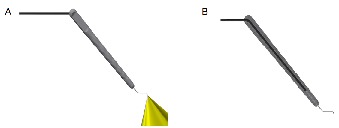
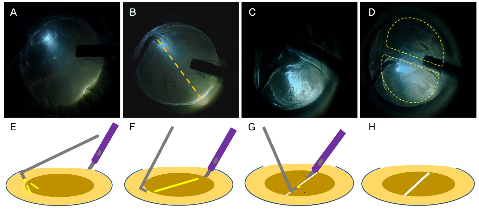
I-Chop은 조명차퍼를 이용한 변형된 double chop 방법으로, 수정체유화술 단계에서 I-chopper를 핵의 적도부에 광원을 이용하여 정확히 고정한 후 수정체유화기와 함께 물리적으로 핵을 조각내는 방식이다[6]. 조명차퍼는 수정체 내의 조명을 통해 수정체 구조물을 더욱 정확히 파악하게 해주어 수정체핵을 물리적으로 더욱 쉽고 안전하게 조각낼 수 있게 하며, 이 단계에서 사용되는 초음파 에너지를 최소화할 수 있다(Fig. 1, 2).

Illustration of illuminated chopper. (A) Nam illumination probe with chopper, Korea and USA Food and Drug Administration cleared (Oculight, Seongnam, Korea). (B) Sectional view of I-chopper.

Procedure of illuminated chop in cataract surgery using I-chopper (A-H). Hooking of I-chopper at the equator of lens with increased visibility of lens structures (A, E). Correct hooking shows linear light pathway under the endonucleus (B, F). Cross the I-chopper and phaco hand piece following the light pathway (C, G). Initial I-chop leading to two large nucleus fragments (D, H).
백내장수술 1달 후 최대교정시력, 안압, 각막내피세포수를 측정하였으며 각막내피세포 감소율(%, [수술 전 각막내 피세포수-수술 1달 후 각막내피세포수]/수술 전 각막내피 세포수)을 구하였다. Stop & Chop 그룹과 I-Chop 그룹 간의 EFX (parameter of effective phaco time with a specific coefficient for the transversal movement expressed in seconds 유효 초음파 에너지양을 나타내는 수술 기구의 고유 상수), 각막내피세포 감소율, 수정체유화술 시간 등을 비교하였으며 I-Chop 그룹에서 EFX=0인 군과 EFX>1인 그룹으로 세분화하여 추가적으로 분석하였다. EFX=0인 군은 수정체유화술 단계에서 유의미한 초음파 에너지의 사용 없이 물리적인 힘과 vacuum으로 수정체유화술이 진행되었음을 의미하며, EFX>1인 군은 초음파 에너지가 사용된 그룹이었다. 통계학적인 분석은 SPSS 18.0 (IBM Corp., Armonk, NY, USA)을 이용하였으며, 두 그룹 간 평균 비교는 independent t-test, Mann-Whitney test를 이용하였고, 모든 통계학적 유의성은 p<0.05로 정의하였다.
결과
대상 인원은 총 83명으로 남자 38명, 여자 45명으로 평균 나이는 65.63 ± 8.03이었다. Stop & Chop 그룹은 34명 (45안)이었으며 I-Chop 그룹은 총 49명(71안)이었다. 두 그룹 간의 나이, 성별, 술 전 안구 계측치는 유의미한 차이가 없었다(Table 1).
I-Chop 그룹에서 EFX는 0.82 ± 3.53, I-Chop을 시행하지 않은 그룹에서는 18.08 ± 16.15였고, 수정체유화술이 이루어진 시간, surgical phaco time은 각각 162.04 ± 49.65, 185.08 ±41.42였으며 두 그룹 간 통계적으로 유의한 차이를 보였다(p=0.001, p=0.01, Table 2).

Comparison of EFX, surgical phaco time, endothelial cell loss, best corrected visual acuity between Stop & Chop group & I-Chop group
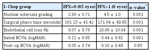
두 그룹 간 술 전 시력(logMAR)은 각각 0.14 ± 0.56, 0.14 ± 0.60, 술 후 시력은 각각 logMAR 0.06 ± 0.65, 0.06 ± 0.69였으며 각막내피세포 감소량은 각각 7.03 ± 7.89, 7.13 ± 9.47%였으며, 두 그룹 간 통계적으로 유의미한 차이를 보이지 않았다(p=0.87, p=0.83, p=0.76, Table 2). 전체 수정체유화술 단계에서 EFX=0인 그룹의 비율을 분석하여 보았을 때, I-Chop 그룹에서는 91.55% (65안)가 EFX=0였으며 Stop & Chop 그룹에서는 EFX=0인 환자는 없었다. I-Chop을 사용한 그룹에서 EFX=0인 군과 EFX>1인 군을 비교하였을 때, 두 그룹 간 각막내피세포감소량은 각각 6.07 ± 8.76, 18.06 ± 10.04, surgical phaco time은 161.13 ± 41.42, 171.04 ± 49.65, 수정체핵경화도는 2.90 ± 0.71, 4.5 ± 1.0으로 모두 통계적으로 유의미한 차이를 보였다 (p=0.001, p=0.001, p=0.01, Table 3).
고찰
백내장수술에서 수정체유화술 중 수정체핵을 완전하게 조각내는 것은 안전한 수술을 위한 중요한 단계이다. 핵을 제거하는 과정에 초음파를 이용하는 것이 가능해짐에 따라 수정체 초음파유화술은 지난 25년간 가장 안전하고 효과적인 수술 방식으로 자리 잡았다. 이 방법은 이전 고식적인 백내장수술보다 안전하고 합병증도 낮은 것으로 알려져 있지만, 높은 초음파 에너지의 사용과 긴 초음파 에너지 사용 시간은 특별한 안과적 과거력이 없는 환자에서 각막내피세포의 감소와 깊은 연관이 있는 것으로 알려져 있다[7]. 더 나아가 높은 초음파 에너지는 백내장수술 후 낭포황반부종의 발생률도 높이기 때문에 수술에서 사용되는 에너지를 최소화하는 것은 환자의 술 후 예후에 중요하다[8].
초음파 에너지는 수정체유화술 단계에서 가장 많이 사용되며, 중등도 이상의 경도를 가진 핵을 제거하기 위해 흔히 사용되는 수술 기법은 phaco chop과 Stop & Chop으로 알려져 있다. Phaco chop은 수정체 핵의 중심부에 초음파 에너지를 이용하여 유화기 첨단부를 고정한 후 물리적으로 핵을 4조각내는 방식이며, Stop & Chop은 초음파 에너지를 이용하여 수정체 핵에 고랑을 만든 후, 차퍼와 수정체유화기의 첨단부를 이용하여 물리적으로 핵을 조각내는데, 2가지 방식 모두 초음파 에너지가 필수적으로 사용된다[9].
위에 소개된 방식과 달리 Yao et al에 의한 소개된 power-free chop, 변형된 phaco chop은 초음파 에너지 사용량을 최소화하여 수정체핵을 제거하는 한층 더 발달된 수술 기법이다[10]. Power free chop 혹은 double chop 방식은 차퍼와 수정체유화기를 기존과 다른 위치에 사용하여 초음파 에너지와 진공의 사용을 최소화하고, 차퍼와 수정체유화기의 물리적인 힘만으로 핵을 제거한다. Power-free chop의 핵심 과정은 차퍼를 수정체 핵의 적도부에 완벽하게 고정하여 주변 조직의 손상 없이 핵을 물리적으로 조각내는 것인데 이 과정은 learning curve가 존재하며, 차퍼를 고정하는 과정에서 섬모체소대의 손상 혹은 수정체낭파열 등이 발생할 위험성이 있다[6].
본 연구진은 이전 논문에서 조명차퍼가 백내장수술 과정에서 안구 내 구조물의 입체감과 시인성을 향상시켜 후낭 파열과 같은 합병증에서 더욱 자유롭고 안전한 백내장수술이 가능함을 보여주었다. 또한 조명차퍼는 백내장수술을 시작하는 초심자에게서 그 유용성과 안정성을 입증하였으며 이를 이용하여 수정체유화술 단계에서 초음파 사용량을 줄이고자 하였다[11-16].
I-Chop 그룹은 Stop & Chop 그룹에 비해 통계적으로 유의미한 EFX 감소를 보여주었다. I-Chop을 시행하는 과정에서 조명차퍼가 수정체의 구조물들을 선명히 시각화하여, 술자가 차퍼를 핵의 적도부에 위치시키는 상대적으로 어려운 단계를 쉽게 시행하였다. 이후 핵을 물리적인 차핑만으로 쉽게 조각낼 수 있었기 때문에 초음파 에너지를 거의 사용하지 않을 수 있었다. 수술 도중 후낭파열, 섬모체소대손상과 같은 합병증은 발생하지 않았으며, Stop & Chop 그룹에서는 groove를 형성할 때 초음파 에너지의 사용이 필수적이기 때문에, 모든 수술에서 EFX>1인 값을 보였다. 그럼에도 불구하고 stop and chop 방식에서 측정된 EFX 양은 이전 다른 연구들에 비해 높지 않은 값을 보였다[17].
I-Chop 그룹에서 EFX=0인 비율이 약 94%로 굉장히 높은 퍼센트를 보였으며 EFX=0인 그룹에 비해 EFX>1이었던 그룹에서는 상대적으로 Lens Opacity Classification System 분류에 따른 수정체 핵의 경화도가 4.50 ± 1.0으로 핵이 더욱 단단하였다. 이로 인해 상대적으로 진행된 백내장수술의 경우 I-Chop을 사용하더라도 물리적인 힘만으로 핵을 완전히 조각내는 것이 어렵기 때문에 상대적으로 초음파 에너지가 더 사용되었을 것으로 예상된다. 또한 I-Chop 그룹은 Stop & Chop 그룹에 비해 통계적으로 유의미한 짧은 수술 시간을 보여 주었는데, 핵을 물리적으로 조각내는 과정이 초음파 에너지를 이용하는 과정보다 빠르며 조명차퍼가 수정체와 주변 구조물을 시각화하여 술자가 더 안정감 있게 수술이 가능했기 때문이다.
두 그룹 간의 각막내피세포수 감소율이나 수술 후 최대 교정시력은 통계적으로 유의미한 차이를 보이지 않았다. 각막내피세포수와 최대교정시력의 차이가 없었던 이유로는 Stop & Chop 그룹에서의 평균 EFX 값도 다른 논문들에 비해 높지 않았고 상대적으로 핵경화 정도가 덜 진행된 백내장의 수술이었으며 수술 중 사용되는 초음파 에너지를 최소화할 수 있는 숙달된 집도의였던 점을 꼽을 수 있겠다.
본 연구의 제한점으로는 동일 술자에 의한 후향적인 연구 특성상 수정체유화술 기법을 통일하지 못하였던 점을 들 수 있으며 이러한 제한점은 향후 동일 기법을 적용한 전향적 연구를 통해 보완할 수 있을 것이다. 본 연구의 결과는 최소 침습적인 백내장수술에 대한 연구가 끝없이 이루어지는 현 시점에서 시도되지 않은 새로운 수정체유화술 방법을 통해 수정체유화술에 사용되는 초음파 에너지를 크게 감소시킬 가능성을 확인한 것에 의의가 있다. 또한 조명 차퍼를 통해 초심자나 각막혼탁, 소동공 등으로 수술 시야가 나쁜 환자에게 도움이 될 것으로 기대된다.
Acknowledgements
This work was supported by the Korea Medical Device Development Fund grant funded by the Korea government (the Ministry of Science and ICT, the Ministry of Trade, Industry and Energy, the Ministry of Health & Welfare, the Ministry of Food and Drug Safety) (Project Number: 1711138951, KMDF_PR_20200901_0296).
Notes
Conflict of Interest
Dong Heun Nam is the CEO, Oculight Co. The others have no financial conflicts of Interest.
References
Biography
김진수 / Jinsoo Kim
가천대학교 길병원 안과
Department of Ophthalmology, Gachon University Gil Medical Center


