실내 조명 아래에서 스마트폰 사용 시 앞방각 모양 및 안압의 변화
Changes in Iridocorneal Angle Configuration and Intraocular Pressure during Smartphone Use under Room Light
Article information
Abstract
목적
실내 조명에서 스마트폰 사용 시에 안압과 앞방각 모양의 변화를 확인하였다.
대상과 방법
안과적 이상이 없는 19-35세 사이의 건강한 성인을 대상으로 스마트폰 사용 전에 리바운드 안압계로 안압을 측정하고 전안부빛간섭단층촬영기를 이용해 앞방각 이미지를 촬영하였다. 이후 스마트폰을 사용하며 2, 4, 6분에 리바운드안압계로 안압을 측정하고 6분에는 앞방각 이미지도 함께 촬영하였다. 6분 후 스마트폰 사용을 중단하고 2분간 휴식을 취하였다. 8분에는 안압 측정만을 시행하였다.
결과
36명의 대상자가 참여하였으며 스마트폰 사용 전의 안압과 비교했을 때 스마트폰 사용 후에 2분째부터 의미 있게 안압이 증가하였고 4, 6분째에도 안압이 지속적으로 증가하였다(p<0.001). 스마트폰의 사용을 종료하고 2분간 휴식 후 8분째에 안압은 스마트폰 사용 전과 유의한 차이가 없었다(p=1.00). 스마트폰 사용 후 앞방 깊이는 감소하였으나(p<0.05) 앞방각 넓이는 넓어졌다(p<0.05).
결론
실내 조명에서 스마트폰 사용 시 안압이 상승하지만 이는 앞방각의 변화와 상관이 없다. 녹내장 발생의 위험이 있는 환자나 녹내장 진행에 대해 주의가 필요한 환자에서는 이에 대한 주지가 필요할 것으로 생각된다.
Trans Abstract
Purpose
To investigate changes in intraocular pressure (IOP) and iridocorneal angle (ICA) configuration during smartphone use under room light.
Methods
We included healthy adults aged 19-35 years with no ophthalmological abnormalities. All read text on a smartphone for 6 minutes under room light. IOP was measured via rebound tonometry at baseline and at 2, 4, and 6 minutes. ICA images were obtained via anterior segment optical coherence tomography after each IOP measurement. After 6 minutes, participants stopped reading text and rested for 2 minutes. IOP was then measured again.
Results
The IOP significantly increased at 2, 4, and 6 minutes of reading compared to baseline (p < 0.001) but recovered to baseline after 2 minutes of rest (p = 1.000). The anterior chamber depth decreased significantly, and the anterior chamber angle width increased after 6 minutes of smartphone reading (both p < 0.05).
Conclusions
IOP increased when reading smartphone text under room light but the ICA did not change. Prolonged smartphone reading is inappropriate for a patient at risk of glaucoma or glaucoma progression. Such patients should be cautioned.
미국의 설문조사기관에 따르면 현대사회에서 컴퓨터, 태블릿, 스마트폰 등의 전자기기 보급률이 시간이 지날수록 증가하고 있다[1]. 특히 한국의 경우 2018년 휴대폰 보급률은 100%로 전 세계 최고 수준이고 이 중 스마트폰은 95%의 비율을 차지하고 있으며 이용 시간 역시 가파르게 증가하는 추세이다[1]. 스마트폰의 사용 시간이 늘어나면서 다른 안증상들이 나타나는데 미국 검안학회(American Optometric Association)에서는 전자기기의 사용 시에 발생하는 눈 긴장, 따가움, 그리고 시야흐림과 같은 증상들로 구성되는 증후군을 컴퓨터시각증후군(computer vision syndrome)으로 정의하였다[2]. 또한 스마트폰의 장시간 사용 시에 안압상승의 위험성이 있다는 연구 결과들이 보고되었다[3,4]. Ha et al [3]은 밝은 환경과 어두운 환경에서 스마트폰을 사용할 때 안압이 상승함을 보고하였다. Yan et al [4]은 진행하는 근시안을 가진 대상자에서 0 diopters (D)에서 +6.0 D까지 조절력을 가하였을 때 약 1 mmHg의 일시적인 안압상승이 발생하는 것을 보고하였다. Daniel et al [5]은 건강한 소아에서 눈이 조절을 하고 있을 때 쉴렘관의 면적과 앞방각 구조의 변화가 발생하는 것을 관찰하였다. 눈의 조절과 관련하여 그동안 많은 연구가 있었지만 스마트폰 사용 시에 안압상승과 앞방각 구조의 변화를 함께 관찰한 연구는 보고된 바가 없다. 본 연구에서는 스마트폰을 사용할 때 시간에 따른 안압의 변화와 앞방각 모양의 변화 및 앞방각 관련 수치의 변화를 관찰하고 분석하였다.
대상과 방법
본 연구는 2021년 1월부터 2021년 3월까지 경북대학교병원 안과에 내원한 환자 중에 나이가 19-35세 사이이고, 33 cm 거리에서 스넬렌시력표로 측정한 근거리 나안시력이 20/20을 만족하는 참가자들을 대상으로 시행한 연구이다. 녹내장을 포함한 안질환의 병력이 있거나 이전에 안과 수술을 한 경우, 안외상의 기왕력이 있는 경우, 안압이 21 mmHg 이상인 경우와 안압에 영향을 미치는 전신약물을 복용하는 경우에는 제외하였다. 본 연구는 경북대학교병원의 임상연구심의위원회(Institutional Review Board, IRB)의 승인(IRB 승인 번호: 202104030) 아래 진행하였고 헬싱키선언(Declaration of Helsinki)을 준수하였다. 모든 참가자들에게 실험 참가 전에 문서화된 동의서를 배부하여 실험의 세부 사항을 안내한 후 서명을 받았다. 연구 시작 전에 근거리 및 원거리 최대교정시력, 안압, 세극등현미경검사, 산동 후 안저검사를 시행하였다.
안압 및 앞방각의 측정
연구에 사용된 스마트폰(Samsung Galaxy S8, Samsung Electronics Co., Ltd., Seoul, Korea)은 모두 동일하였다. 밝기는 100%로 설정하였고, 기본 탑재된 어플리케이션에서 폰트의 크기는 15로 일괄 설정된 한글 텍스트를 읽었다. 글자의 색깔은 검정색이었고 배경색은 흰색으로 가장 대비되는 색을 이용하였다. 안압 측정은 iCare ic200 (iCare Finland Oy, Helsinki, Finland)를 이용하였다. 앞방각 이미지는 전안부빛간섭단층촬영기(anterior segment optical coherence tomography)인 CASIA SS-1000 (Tomey Corp., Nagoya, Japan)을 사용하여 획득하였다. 연구를 진행하는 공간의 밝기는 142 lux의 실내 조명이었다. 안압 측정 및 앞방각 이미지 촬영은 비우세안에 시행하였으며 실험 시작 전 로젠바흐검사를 시행하여 우세안과 비우세안을 확인하였다.
모든 참가자들은 실험 전날 6시간 이상의 충분한 수면을 취할 수 있도록 하였고 실험 전에 안압이 상승할 수 있는 카페인이 포함된 음료수나 음식은 섭취하지 않도록 하였다[6]. 실험을 시행하기 전에 30분간 앉아서 안정을 취하였으며, 안압의 일중 변동에 대한 변수를 통제하기 위해 모든 실험은 오전 11시에 시작되었다. ic200에 내장되어 있는 연속 측정 모드를 사용하여 6회의 안압을 측정하였다. 6회의 안압 측정 후 자동적으로 가장 높은 측정값과 가장 낮은 측정값을 제외하고 남은 4개의 측정값의 평균값이 화면에 출력되어 이 값을 최종 안압으로 기록하였다[7]. 30분의 휴식 후에 스마트폰 사용 전에 안압을 측정하고 즉시 전안부빛간섭단층촬영기를 이용하여 앞방각 이미지를 촬영하였다. 안압 측정 및 앞방각 이미지의 촬영이 끝나면 의자에 앉은 채로 허리를 곧게 펴고 참가자의 앞에 위치한 책상 위 33 cm 거리에 스마트폰을 위치시킨 후 불편한 부분이 없는지 확인하고 실험용 텍스트를 읽기 시작했다. 2분, 4분, 6분째 비우세안의 안압을 측정하였고 이 때는 눈과 스마트폰의 거리를 33 cm로 일정하게 유지한 채로 고개와 스마트폰을 들어 올려 시선이 지면에 수평이 되도록 자세를 취한 뒤 안압을 측정하였다. 안압 측정 중에도 스마트폰 화면을 주시하며 화면의 텍스트를 계속 읽도록 하였다. 6분째에 안압을 측정한 후 즉시 앞방각 이미지를 촬영하였다. 이때 스마트폰을 사용하여 발생하는 조절이 유사하게 유지되도록 CASIA 탑재 프로그램에서 -3.0 D의 조절력을 설정해 촬영하였으며 조도 환경은 안압측정 시와 동일한 142 lux였다. 그 후 스마트폰 사용을 중지하고 편안한 자세로 앉아 2분간 휴식을 취한 후 다시 안압을 측정하였다.
앞방각은 CASIA 기기(CASIA SS-1000, Tomey Corp.)로 촬영하였고 앞방각 인자들은 기기에 기본 탑재된 소프트웨어에서 반자동으로 인식되어 측정되었으며 이측 부분의 3시 혹은 9시 방향의 앞방각 구조물을 대상으로 분석을 시행하였다. 앞방각 인자는 앞방깊이(anterior chamber depth, ACD), angle opening distance (AOD), angle recess area (ARA), trabecular iris surface area (TISA), trabecular iris angle (TIA), 쉴렘관 면적(Schlemn’s canal area, SCA), 쉴렘관 둘레(Schlemn’s canal circumference, SCC)를 분석하였다(Fig. 1). CASIA에서 관찰되는 쉴렘관의 위치는 이전 연구를 참고하였으며[8,9] 쉴렘관 둘레와 면적은 CASIA에 기본 탑재되어 있는 소프트웨어의 자유 그리기 도구를 이용하여 자동적으로 계산하였다. 스마트폰 사용 전과 6분째 촬영하여 획득한 앞방각 인자들과 SCA, SCC를 비교 분석하였다.
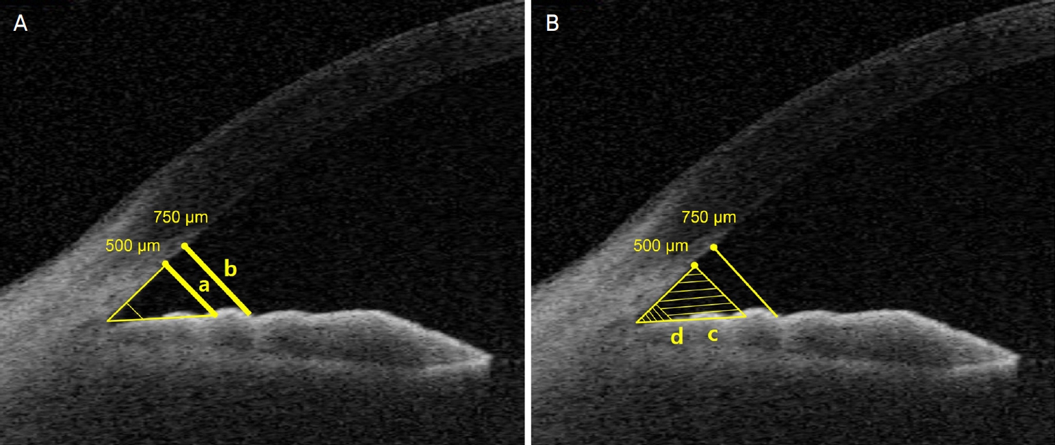
(A) AOD500 (a) is the distance between the posterior cornea surface and the anterior iris measured on a line perpendicular to the trabecular meshwork at 500 μm from the scleral spur (SS). AOD750 (b) is the distance that measured with same manners at 750 μm from the SS. (B) TISA500 (c) is the surface area of a trapezoid with the following boundaries: anteriorly, the perpendicular line between scleral spur and the opposing iris; posteriorly, the AOD500, superiorly, the inner corneal wall; and inferiorly, the iris surface. TIA500 is a trabecular iris angle at 500 μm from the SS. ARA500 (c + d) is the triangular area that demarcated by the trabecular meshwork, anterior iris surface, and corneal endothelium with distance of 500 μm from the SS.
연구에 필요한 전체 연구 대상자 수를 산출하기 위해 G-Power (Version3.1.9.7; Franz Faul, University of Kiel, Kiel, Germany)를 사용하였다. 0.05의 유의수준에서 95%의 검정력을 가지기 위해 최소 31명의 연구 대상자가 필요하였다. 통계 분석은 SPSS Version 26.0 (IBM Corp., Armonk, NY, USA)를 사용하였다. 스마트폰 사용 전과 사용 후 2, 4, 6, 8분의 안압 변화는 반복측정 분산분석(repeated measured analysis of variation)을 사용하여 분석하였고 사후분석으로 Bonferonni’s test를 시행하였다. 스마트폰 사용전과 사용 후 6분의 앞방각 인자의 비교에는 paired t-test를 사용하였다. 통계 결과의 검정에 있어 p값이 0.05 미만일 때 통계적으로 유의한 것으로 간주하였다.
결 과
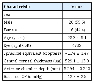
37명의 37안이 등록되었으나 1명의 지원자는 안외상의 과거력이 확인되어 연구에서 배제되어 총 36안에서 연구가 진행되었다. 36명의 참가자 중 남성은 20명(55.6%), 여성은 16명(44.4%)이었다. 평균 나이는 28.3 ± 3.1세였고 우안이 4안, 좌안이 32안이었다. 평균 굴절값은 -1.74 ± 1.47 D였고 평균 중심각막두께는 529.1 ± 13.0 μm, 스마트폰 사용 직전의 안압은 12.7 ± 2.5 mmHg였다(Table 1).
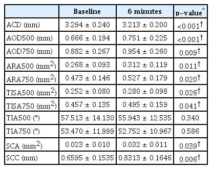
스마트폰 사용 후 2분째부터 유의한 안압상승이 관찰되었고(14.3 ± 2.9 mmHg, +13.2%, p<0.001), 6분째 측정한 안압이 가장 높았다(15.4 ± 3.3 mmHg, +21.5%, p<0.001). 스마트폰 사용 후 2, 4, 6분째의 안압은 스마트폰 사용 직전의 안압에 비해 높게 측정되었다. 6분째 안압 측정을 마치고 2분간 스마트폰 사용을 중단한 후 8분째 측정한 안압은 스마트폰 사용 직전의 안압과 유사한 정도로 하강하였다(12.8 ± 2.5 mmHg, +1.1%, p=1.0) (Fig. 2, 3). 스마트폰을 사용하는 동안 ACD는 감소하였으나 AOD, ARA, TISA 등은 의미 있게 증가하였고, SCA과 SCC도 의미 있게 증가하였으며 TIA는 감소하는 경향이었으나 통계적으로 유의하지는 않았다(Table 2).
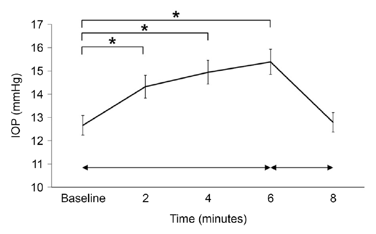
Mean intraocular pressure (IOP) during smartphone reading (error bars represent ± standard error). *Statistically significant by repeated measured analysis of variation (p < 0.001).

Percent change of intraocular pressure (IOP) from baseline during smartphone reading (error bars represent ± standard error). *Statistically significant by repeated measured analysis of variation (p < 0.001).
고 찰
본 연구에서는 실내 조명 하에서 스마트폰을 사용한 후에 안압이 의미 있게 상승하였는데, 이전 연구에서는 근거리 작업 혹은 조절 후에 안압이 증가한다는 연구[3,4,10]와 감소한다는 상반되는 연구 결과[11-13]가 보고되었다. Ha et al [3]은 스마트폰 사용 시에 안압이 증가하는 이유로 조절과 눈모음, 외안근의 수축, 정신적인 스트레스, 건성안, 고개를 숙인 자세 등을 제시하였다. 본 연구에서는 스마트폰으로 조절을 유발한 결과 안압이 빠른 시간 내에 증가하였다. 외안근의 수축과 관련하여 Coleman and Trokel [14]은 좌측으로 볼 때 5-10 mmHg의 안압상승이 발생함을 보고하였다. 이는 안구 내부 요인이 아닌 외부의 외안근이 수축하면서 안압이 상승할 수 있음을 시사하는 것이다. 본 연구에서는, 안압 측정을 수행하기 직전까지 자연스럽게 스마트폰의 텍스트를 읽고, 안압을 측정할 때는 주시안으로는 스마트폰을 지속적으로 주시하며 눈을 움직이지 않고 고정한 채로 비주시안을 리바운드 안압계로 빠르게 검사하여, 외안근의 수축과 안구의 좌우 운동에 의한 안압상승을 최대한 배제하였다. 그러나 스마트폰의 글자를 읽기 위한 미세한 안구운동은 다 제어하지 못했을 것으로 생각된다. Malihi and Sit [15]는 고개를 젖혔을 때보다는 숙였을 때 안압이 증가한다고 보고하였다. 본 연구에서는 눈과 스마트폰의 거리를 33 cm로 일정하게 유지한 채로 고개와 스마트폰을 들어 올려 시선이 지면과 수평이 되도록 자세를 취한 뒤에 안압을 측정하여 고개를 숙였을 때 나타날 수 있는 안압상승을 최대한 배제하고자 하였다.
이러한 안압의 하루 중 변동은 녹내장 환자에서 녹내장의 진행에 중요한 역할을 하는데[16] Ha et al [10]은 약물로 조절되는 정상안압녹내장 환자와 섬유주절제술을 시행한 녹내장 환자에서 스마트폰을 사용할 때에 안압이 의미 있게 증가하는 것으로 보고하였는데 섬유주절제술을 시행한 경우에 안압의 변동이 훨씬 줄어드는 것으로 보고하였다. 본 연구에서는 Ha et al [3,10]의 연구보다 더 빠른 시간(2분)에 안압이 상승하였으며 스마트폰 사용을 중단할 경우에 비교적 빠르게(2분) 이전 안압으로 회복되는 것으로 나타나 스마트폰의 사용과 같은 일상의 활동 중에 안압의 변동이 비교적 빠르게 나타남을 알 수 있었다.
이전의 여러 연구에서 수정체의 조절 시에 ACD는 줄어드는 것으로 보고되었다[4,17-23]. 조절이 일어날 때 ACD가 줄어드는 이유는 조절이 일어나면 수정체가 두꺼워지고 수정체 앞면과 뒷면의 곡률반경이 작아지며 수정체가 앞으로 이동한다[4,17-22]. 본 연구에서도 스마트폰 사용 전과 비교해 6분째 ACD는 줄어들었으나 수정체에 대한 분석은 따로 시행하지 않았다.
본 연구에서 스마트폰 사용 시에 섬유주 주변의 앞방각 구조는 넓어지는 결과를 보였다(Table 2). 이는 근거리 작업 시 조절을 할 때 앞방각 인자들이 커지며 앞방각이 넓어진다는 이전 연구의 결과와도 일치하는 부분이다[5,23,24]. 반면 일부 보고에서는 조절 시에 앞방각의 변화는 관찰되지 않았으며[17,25] 앞방각이 좁아진다는 보고도 있다[4]. 본 연구와 같이 조절 시에 앞방각이 넓어지는 이유에 대해서는 잘 알려지지 않았으나 Martinez-Enriquez et al [26]은 젊은 환자에서 조절 시에 수정체의 적도부 직경이 감소하는 것을 보고하였다. 앞서의 연구들과 더불어 유추한다면 조절 시에는 수정체가 두꺼워지고 수정체 앞면과 뒷면의 곡률반경이 적어지고 수정체의 적도부 직경이 감소한다. 수정체의 두께가 증가하고 적도부 직경이 감소하면 앞방각 근처의 홍채는 수정체쪽으로 휘어지게 되고 앞방각이 넓어지게 된다.
앞방각 폐쇄에 의한 폐쇄각녹내장의 경우 홍채 볼록이 생성되어 섬유주와 홍채가 접촉하여 방수 유출이 되지 않아 안압이 상승하게 된다[8,27]. 하지만 앞방각이 정상인 대상자에서 시행한 본 연구에서는 수정체가 조절함에 따라 ACD는 줄어들지만 앞방각 주변의 홍채는 홍채 볼록이 형성되는 방향의 반대 방향으로 하강하였고 이에 따라 앞방각 구조가 넓어지는 변화를 전안부빛간섭단층촬영으로 확인하였다. 이 결과에 비춰 보았을 때 스마트폰 사용 시에 수정체의 조절에 따른 안압상승은 앞방각 폐쇄의 기전과는 다른 기전이 작용할 것으로 생각된다.
본 연구에서는 리바운드 안압계를 사용하였는데 골드만압평안압계는 녹내장 환자들의 안압 측정에 표준이 되지만[28] 스마트폰을 사용하면서 조절을 유지하는 동안에 골드만압평안압계로 안압을 측정하기에는 준비 과정이 많고 안압을 측정하는 순간 근거리 대상에 대한 집중력을 잃고 조절을 유지하지 못하여 조절에 의한 안압 변화를 정확히 반영하지 못할 수 있다. 리바운드 안압계의 정확도는 7-22 mmHg의 범위에서 골드만압평안압계와 차이가 없다는 연구 결과가 있으며[29] 안압을 측정하는데 사전 준비가 필요 없고 실험 대상자가 우세안으로 주시 대상을 집중하여 보면서 안압을 측정할 수 있는 장점이 있다. 본 연구에서 사용한 리바운드 안압계는 참가자들의 평균적인 안압 분포(7.8-21.4 mmHg)에서 골드만압평안압계 만큼이나 정확한 안압을 측정할 수 있을 것으로 판단하였다.
본 연구에는 몇 가지 제한점이 있다. 첫째, 연구 대상의 수가 적고 35세 이하의 젊은 지원자로 제한하였으며, 대부분의 경우 정시안으로 원시나 백내장 혹은 녹내장 환자들이 포함되지 않았다. 후속 연구에서는 나이 또는 조절근점정도에 따라 실험군을 달리 설정하고 이들 군 간의 비교를 시행하여 안압상승에 영향을 미치는 요인별 분석이 필요할 것이다. 둘째, 수정체 천장높이(lens vault)와 안축장(axial length)에 의해서 앞방각의 모양과 넓이가 영향을 받을 수 있기 때문에 이에 따른 분석이 필요하다. 셋째, 실제로 스마트폰 사용 중에 안압이 상승하는 것이 녹내장의 발생 또는 진행에 영향을 미치는지에 대한 연구가 필요하다. 장기간의 추적 관찰을 하여 스마트폰을 사용하는 정도의 일상적 활동에 의해 정상인에서 녹내장이 발생하는 것을 관찰하거나, 녹내장 환자에서 녹내장이 진행하는지에 대한 장기적인 연구가 필요하다.
본 연구에서는 실내 조명 아래에서 스마트폰을 사용할 때 안압상승이 발생하는 것을 확인하였다. 앞방각의 변화와는 상관이 적어 보이지만 녹내장에 대한 경과 관찰이 필요하거나, 녹내장 진행에 대한 주의가 필요한 환자들에게는 스마트폰의 근거리 사용이 위험요소가 될 수도 있으니 이에 대한 주지가 필요할 것으로 생각된다.
Notes
DHP is financially supported by the Basic Science Research Program of the National Research Foundation of Korea (NRF), funded by the Korean government (Ministry of Science and ICT) (2019R1A2C1084371), and the Ministry of Science and ICT (MSIT), Korea, under the Information Technology Research Center (ITRC) support program (IITP-2021-2020-0-01808) supervised by the Institute of Information & Communications Technology Planning & Evaluation (IITP).
References
Biography
이성택 / Seong Taik Lee
경북대학교 의과대학 경북대학교병원 안과학교실
Department of Ophthalmology, Kyungpook National University Hospital, School of Medicine, Kyungpook National University
