중심장액맥락망막병증에서 유리체강내 베바시주맙과 애플리버셉트 주입술의 비교
Comparison of Intravitreal Bevacizumab and Aflibercept Injections for Central Serous Chorioretinopathy
Article information
Abstract
목적
중심장액맥락망막병증 환자에서 유리체강내 베바시주맙 주입술과 애플리버셉트 주입술 간의 치료 효과 차이를 알아보고자 한다.
대상과 방법
처음 중심장액맥락망막병증으로 진단받고 유리체강내 항혈관내피성장인자 주사를 받은 환자 49명 51안을 대상으로 하여 의무기록을 후향적으로 분석하였다. 항혈관내피성장인자 주사 종류에 따라 유리체강내 베바시주맙을 주입한 군과 애플리버셉트를 주입한 군으로 구분하였고, 시술에 대한 반응이 없거나 악화 시 동일한 약제로 반복 치료하였다. 치료 이후 3개월 이상의 추적 관찰을 시행하였고, 최대교정시력과 중심맥락막두께 변화, 치료까지 시행된 주사 횟수 및 기간을 분석하였다.
결과
유리체강내 베바시주맙 주입술과 애플리버셉트 주입술 모두 치료 전후 의미 있는 시력변화(p<0.0001, p=0.001)와 중심맥락막 두께 변화를 보였다(p<0.0001, p=0.011). 하지만 주입 약제에 따른 치료 전후 최대교정시력 및 중심맥락막두께 변화량의 차이는 보이지 않았다. 또한 주입 약제에 따른 치료까지 시행된 주사 횟수 및 기간도 차이를 보이지 않았다.
결론
중심장액맥락망막병증 환자에서 유리체강내 베바시주맙 주입술 또는 애플리버셉트 주입술 모두 중심장액맥락망막병증의 구조적 및 기능적 회복에 효과가 있었으나, 약제의 종류에 따른 기능적 그리고 해부학적 호전의 정도, 치료까지 시행된 주사 횟수 및 기간의 차이는 없었다.
Trans Abstract
Purpose
We examined differences in the treatment effects of intravitreal bevacizumab injections and intravitreal aflibercept injections in patients with central serous chorioretinopathy.
Methods
This retrospective analysis included 51 eyes of 49 patients who received intravitreal anti-vascular endothelial growth factor agent injections after initial diagnosis with central serous chorioretinopathy. The patients were divided into two groups: one received an intravitreal bevacizumab injection, and another one received an intravitreal aflibercept injection. Patients with no reaction to treatment or a worsened condition, received repeat treatment with the same therapy. After treatment, patients were monitored for >3 months. Data were collected regarding best- corrected visual acuity (BCVA), subfoveal choroidal thickness, injection number, and treatment duration.
Results
Both intravitreal bevacizumab injections and intravitreal aflibercept injections led to significant differences in BCVA (p < 0.0001, p = 0.001) and subfoveal choroidal thickness (p < 0.0001, p = 0.011), compared between before and after treatment. However, no differences between groups were observed in mean change of BCVA or subfoveal choroidal thickness. In addition, there were no differences between groups in injection number and treatment duration.
Conclusions
In patients with central serous chorioretinopathy, both intravitreal bevacizumab injections and intravitreal aflibercept injections are effective treatment methods. There were no differences between the two medicines in terms of functional and anatomical recovery, or the injection number and treatment duration.
중심장액맥락망막병증(central serous chorioretinopathy)은 맥락막모세혈관으로부터 누출된 삼출물로 인한 황반부의 신경망막박리를 특징으로 하는 질병이다[1]. 정확한 병인과 병태생리는 밝혀져 있지 않으나 맥락막모세혈관의 비정상적인 투과성 증가와 망막색소상피의 기능 저하에 의해 황반부의 망막 아래(sub-retina)에 장액성 액체가 축적되어 발생하는 것으로 알려져 있다[2]. 급성 중심장액맥락망막병증의 경우 일반적으로 자연 호전되는 경과를 보이지만 1년 내에 재발하는 경우가 많다[2]. 임상적으로 사용되는 치료 방법으로는 광역학치료가 있는데 이는 망막색소상피의 위축, 맥락막모세혈관 허혈, 황반주변부의 이차 맥락막신생혈관 발생 등의 합병증들이 보고되었다[3]. 때문에 구조적인 손상을 최소화하면서, 맥락막혈관의 과투과성을 유발하는 혈관내피성장인자(vascular endothelial growth factor, VEGF)를 억제하는 항혈관내피성장인자(anti-VEGF agent) 치료가 고려되었으며[4], 최근 여러 연구들에서 중심망막두께의 감소, 최대교정시력 개선 등의 효과적인 결과를 보였다[5-11].
Anti-VEGF agent에서 베바시주맙(AvastinⓇ, Genentech, South San Francisco, CA, USA)은 VEGF-A를 억제하여 작용하지만[12,13], 이후 개발된 애플리버셉트(EyleaⓇ, Regeneron Pharmaceuticals, Inc, Tarrytown, NY, USA; Bayer Pharma AG, Berlin, Germany)는 VEGF-A뿐 아닌 VEGF-B와 태반성장인자(placental growth factor)에도 결합하여 작용하는 특징[14]과 VEGF-A에 더 빠르게 결합하며 100배 높은 결합력을 지닌 장점을 가졌다[15-18]. 그러나 국내에서는 중심장액맥락망막병증이 애플리버셉트의 보험적용 대상 질환이 아니기에, 사용 시 환자가 약값을 전부 부담해야 하는 경제적인 요인으로 인하여 중심장액맥락망막병증 환자에서 유리체강내 애플리버셉트 주입술을 시행하기 쉬운 여건이 아니다. 또한 국내외에서 중심장액맥락망막병증에서 두 약제를 비교한 연구는 드물다. 이에 본 연구에서는 중심장액맥락망막병증 환자에서 약제 종류에 따른 시력 및 중심맥락막 두께의 변화, 치료에 필요한 기간 및 주사 횟수 등을 분석함으로써, 유리체강내 베바시주맙 주입술과 애플리버셉트 주입술의 효과를 비교해 보고자 하였다.
대상과 방법
2010년 1월부터 2020년 7월까지 본원에서 처음 중심장액맥락망막병증으로 진단받고 최소 3개월 동안 시력과 황반부의 안정된 소견을 보일 때까지 유리체강내 베바시주맙주입술 혹은 애플리버셉트 주입술을 시행 받은 치료군(treatment group) 총 49명 51안의 의무기록을 후향적으로 분석하였다. 다음 기준을 충족하는 경우 연구에 포함하였다: 1) 스펙트럼 도메인 빛간섭단층촬영(Spectralis OCTⓇ, Heidelberg Engineering, Heidelberg, Germany)에서 명확하게 망막하액(subretinal fluid, SRF)이 관찰되는 경우, 2) 형광안저혈관조영술(Spectralis HRA2Ⓡ, Heidelberg Engineering, Heidelberg, Germany)에서 망막하액과 연관된 누출점이 관찰되는 경우, 3) 마지막 안구내주사 후 3개월 이상의 경과 관찰이 가능한 경우. 다음과 같은 경우는 연구에서 제외하였다: 1) 맥락막신생혈관(choroidal neovascularization, CNV), 연령관련환반변성 등 다른 황반질환이 있는 경우, 2) 주사 치료 기간 중 유리체강내 트리암시놀론 주입술, 광역학치료, 망막광응고술 등 다른 시술을 받은 경우, 3) 주사치료 기간 중 유리체절제술, 백내장수술 등 다른 수술을 한 경우, 4) 동일 약제를 사용하지 않고 중간에 베바시주맙에서 애플리버셉트 혹은 애플리버셉트에서 베바시주맙 같이 약제가 바뀐 경우. 본 연구는 헬싱키선언을 준수하였으며, 본원 임상연구윤리심의위원회(Institutional Review Board, IRB)의 승인 하에 진행되었다(IRB 승인 번호: 2021-01-006-001).
환자가 처음 외래를 내원하였을 때 스넬렌시력표를 이용한 최대교정시력 측정, 비접촉안압계(KT-800Ⓡ, Kowa American Corp., New York, NY, USA)를 이용한 안압 측정, 안저검사, 스펙트럼 도메인 빛간섭단층촬영, 형광안저혈관조영술이 시행되었다. 진단 후 1주일 이내에 유리체강내 베바시주맙 주입술 혹은 애플리버셉트 주입술을 시행하였다. 안구 내 주사 전에 모든 환자에게 경과 관찰, 광역학치료 등 다른 치료 방법들을 설명하였고, 주사 약제들에 대한 충분한 설명과 함께 환자가 직접 약제를 선택하도록 하였으며, 또한 중심장액맥락망막병증에서 허가초과용제로 사용되는 약제임을 설명하고 사전 동의를 받았다.
주사 후 1일째 외래를 방문하여 안압상승 여부 및 안내 염증 발생 유무 등의 합병증 여부를 확인하였으며, 약 1개월 간격으로 추적 관찰을 시행하였다. 매 회의 추적 관찰에서 최대교정시력과 중심맥락막두께 측정이 이루어졌다. 본 연구에서 ‘치료’는 3개월 이상의 경과 관찰에서 빛간섭단층촬영상 SRF가 사라진 것으로 정의하였다. 추적 관찰 중 빛간섭단층촬영에서 망막하액의 지속 혹은 증가가 확인되는 경우 주사치료에 반응이 없거나 악화가 되는 것으로 간주하였으며 추가 주사치료가 시행되었다.
통계적 분석을 위해 최대교정시력은 logarithm of the minimal angle of resolution (logMAR) scale로 변환하였다. 중심맥락막두께는 빛간섭단층촬영 기기의 디지털 캘리퍼기능을 사용하여 중심와 위치에서 망막색소상피로부터 맥락막-공막 안쪽 경계까지의 거리를 수직으로 측정하였다. 환자는 베바시주맙 주입술을 시행 받은 군(베바시주맙 군)과 애플리버셉트 주입술을 시행 받은 군(애플리버셉트 군) 두 군으로 나누었다. 치료 전과 치료 후 3개월째의 최대교정시력, 중심맥락막두께를 비교하였으며, 두 군 간 최대교정시력과 중심맥락막두께의 변화량 및 시행된 주사 횟수, 치료까지의 기간을 비교하였다.
데이터분석에는 SPSS Statistics 18 (IBM, Armonk, NY, USA) 소프트웨어를 이용하였다. 본 연구는 표본수가 적어 모집단의 정규성 검토를 위하여 Kolmogorov-Smirnov의 정규성 검정을 실시하였다. 정규성을 보이는 두 군 간의 비교는 independent t-test를, 정규성을 보이지 않는 두 군은 Mann-Whitney test를 통하여 검정하였다. 정규성을 보이는 전후 비교는 paired t-test를, 정규성을 보이지 않는 전후 비교는 Wilcoxon signed rank test를 사용하였다. 명목척도의 자료 분석에는 chi-squared test를 통하여 검정하였다. 모든 결과값은 p값이 0.05 미만일 경우 통계적으로 유의한 것으로 간주하였다.
결 과
전체 51안(49명) 중 베바시주맙 군이 32안, 애플리버셉트 군이 19안이었다. 베바시주맙군에서는 남자 18안(56.3%)으로 평균연령은 50.44 ± 7.14세였고, 애플리버셉트군에서는 남자 15안(78.9%)으로 평균연령은 55.79 ± 12.39세였다. 치료 전 환자군의 특성은 Table 1에 나타나 있다.
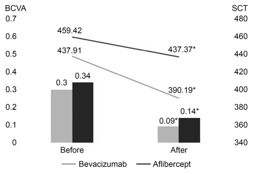
베바시주맙 군의 최대교정시력은 치료 전 0.30 ± 0.22에서 치료 후 0.09 ± 0.10으로, 애플리버셉트 군은 치료 전 0.34 ± 0.23에서 치료 후 0.14 ± 0.16으로 통계적으로 의미 있게 좋아졌으며(각각, p<0.0001, p=0.001), 중심맥락막두께(μm) 또한 베바시주맙 군에서 치료 전 437.9 1 ± 79.72에서 치료 후 390.19 ± 74.72로, 애플리버셉트 군은 치료 전 459.42 ± 45.14에서 치료 후 437.37 ± 37.38로 통계적으로 의미 있게 감소하였다(각각, p<0.0001, p=0.011) (Fig. 1).

Comparison of best-corrected visual acuity (BCVA) and subfoveal choroidal thickness (SCT) according to the group of injection type before and 3 months after treatment. Gray bar and gray line mean bevacizumab group, Black bar and black line mean aflibercept group. *Statistically significance is p < 0.05 with baseline.
베바시주맙와 애플리버셉트 두 군 간 치료 전후 분석에서 최대교정시력의 변화량은 베바시주맙군에서 0.21 ± 0.21, 애플리버셉트 군에서 0.20 ± 0.17이었고, 중심맥락막두께 변화량은 베바시주맙 군에서 47.72 ± 57.78, 애플리버셉트 군에서 22.05 ± 30.36이었으나 통계적으로 유의한 차이는 없어, 베바시주맙 주입술과 애플리버셉트 주입술 간의 기능과 해부학적 회복의 차이를 보이지 않았다(Table 2).
치료까지 걸린 기간은 베바시주맙 군이 101.97 ± 100.42일, 애플리버셉트 군이 80.74 ± 75.80일이 걸렸으나 통계적으로 유의한 차이는 없었다(p=0.453). 1회 주사치료 이후 호전되지 않아 추가 치료를 한 경우는 베바시주맙 군에서 32안 중 총 7안이었으며, 그 중 3안(9.38%)이 2회, 4안(12.5%)이 3회 주사를 시행하였다. 애플리버셉트 군은 19안 중 총 6안이 추가 치료를 시행 받았으며, 그 중 4안(21.05%)이 2회, 1안(5.26%)이 3회, 1안(5.26%)이 4회 주사를 시행 받았다. 결과적으로 치료까지의 주사 횟수는 베바시주맙 군이 1.34 ± 0.70회, 애플리버셉트 군이 1.47 ± 0.84회로 통계적으로 유의한 차이는 없었다(p=0.499). 전체 경과 관찰 기간 동안 51안에 대하여 안압상승, 안내염, 망막박리, 유리체출혈 및 전신적 부작용은 관찰되지 않았다.
고 찰
중심장액맥락망막병증의 병리기전은 영상검사기술의 발전과 수많은 연구들에도 불구하고 여러 가지 가설들이 제시되었을 뿐 명확하게 밝혀지지 않았다. 최근 가장 유력한 가설은 맥락막모세혈관의 투과성 증가이다[19]. 맥락막의 울혈, 허혈 또는 염증 등에 의한 맥락막모세혈관의 과투과성 때문에 맥락막 내 정수압이 증가하고[20], 망막색소상피박리가 촉진되어 감각신경망막과 망막색소상피 사이에 삼출물이 축적된다는 가설이다. 급성 중심장액맥락망막병증은 1-4개월 안에 감각신경망막의 재부착과 함께 시력이 회복되는 자연 호전의 경과를 보이기도 한다[19]. 그러나 30-50% 환자에서 1년 안에 재발이 흔하며, 빈번한 재발은 망막색소상피의 위축과 감각신경망막의 변화를 일으켜 시각기능을 영구히 잃을 수 있기 때문에 광역학치료 및 유리체강내 anti-VEGF 주입술 같은 치료를 시도해왔다.
Anti-VEGF 치료는 중심장액맥락망막병증에서 맥락막과 망막색소상피의 기능 및 구조 변화로 인해 저관류, 저산소상태가 초래되면 혈관내피성장인자(VEGF)가 발현된다는 가설 하에 사용되었으며[21], 유리체강내에 anti-VEGF를 주사하여 VEGF를 억제시키면 맥락막혈관의 과투과성에 변화를 주어 망막하액을 감소시킬 수 있다는 보고가 있다.16,17 급성 중심장액맥락망막병증에서 베바시주맙이나 라니비주맙(LucentisⓇ, Genentech Inc., South San Francisco, CA, USA) 치료군이 경과 관찰군보다 해부학적 호전이 빠르다는 연구 결과들이 있으며[22-24], Koh et al [25]은 베바시주맙 치료가 급성 중심장액맥락망막병증의 재발률을 낮추는 데에 기여한다고 하였다. Jung et al [26]은 급성 중심장액맥락망막병증에서 애플리버셉트 치료군이 의미 있는 중심망막두께와 맥락막두께의 호전을 보였고, 대조군보다 더 빠른 시력 호전을 보였다고 보고하였다. 만성 중심장액맥락망막병증을 대상으로 한 전향적인 연구에서 베바시주맙과 애플리버셉트 치료가 해부학적 및 기능 호전을 보인다는 연구 결과들도 있다[27,28]. 본 연구에서도 유리체강내 anti-VEGF 주사를 초기 치료로 사용한 환자들을 대상으로 연구를 진행하여 베바시주맙군과 애플리버셉트군 모두 합병증 없이 최대 교정시력의 호전과 중심맥락막두께 감소라는 기능적, 해부학적으로 유의한 효과를 관찰할 수 있었다.
중심장액맥락망막병증 외에 대표적인 망막혈관질환인 당뇨망막병증이나 중심망막정맥폐쇄에서는 애플리버셉트가 베바시주맙보다 효과적이라는 연구 결과들이 있다. Wells et al [29]과 Heier et al [30]는 당뇨망막병증에서 애플리버셉트가 베바시주맙을 사용한 군보다 시력호전에 있어 효과적이라고 하였다. Lotfy et al [31]은 중심망막정맥폐쇄에서 애플리버셉트와 베바시주맙 모두 효과가 있었지만, 애플리버셉트를 사용한 군에서 더 긴 주사 간격과 더 적은 횟수의 주사로 효과가 나타났기 때문에 애플리버셉트가 베바시주맙보다 효과적이며, 이는 애플리버셉트의 세포수용체 결합 능력이 더 높아 VEGF를 억제하는 능력이 더 좋기 때문이라고 하였다. 중심장액맥락망막병증에서 두 약제를 비교한 본 연구에서는 베바시주맙 군과 애플리버셉트 군 간의 치료 전 후 최대교정시력 및 중심맥락막두께 변화량을 비교하였을 때 유의한 차이를 보이지 않았다. 이는 앞서 언급한 다른 망막혈관질환들의 연구와는 상이한 결과이다. 본 연구와 비슷하게 급성 중심장액맥락망막병증에서 베바시주맙과 라니비주맙의 효과를 비교 분석한 연구에서도 라니비주맙주사군이 베바시주맙 주사군보다 망막하액의 소실되기까지의 기간이 더 짧게 걸리기는 하였으나 통계적으로 유의하지는 않았다는 결과가 있다[24].
당뇨망막병증이나 망막중심정맥폐쇄에서는 유리체강내 VEGF 농도가 명확하게 높다[32,33]. 하지만 중심장액맥락망막병증 환자에서는 방수 내 VEGF 농도가 정상 대조군과 차이를 보이지 않았다[34,35]. 때문에 높은 결합력을 가진 애플리버셉트의 이점을 상쇄시켰을 것으로 사료된다. Jung et al [26] 또한 중심장액맥락망막병증 환자의 앞방수 농도가 건강한 사람과 비교하였을 때 차이가 없었기 때문에 중심장액맥락망막병증의 치료로 anti-VEGF 약제를 사용할 때 약제의 결합력이 생물학적 활성도를 결정하는 주요 요인이 아니라고 보고하였다. 치료약제의 생물학적 활성도는 약제의 결합력 뿐만 아니라 반감기의 영향을 받는다[36]. 사람의 안구 내에서 애플리버셉트의 반감기에 대한 연구는 아직 없지만, 안구내 반감기는 분자량에 의해 결정되므로 베바시주맙(분자량: 149 kDa)에 비해 분자량이 더 작은 애플리버셉트(분자량: 115 kDa)의 반감기가 더 짧을 것으로 생각된다[37]. 때문에 반감기가 더 길다는 이점을 지닌 베바시주맙이 본 연구에서 애플리버셉트와 비슷한 효과를 보인 것으로 사료된다.
본 연구는 몇 가지 한계점을 지닌다. 우선, 급성과 만성중심장액맥락망막병증 각각의 군에 대하여 치료 약제 간의 효과를 비교 분석하지 못하였다. Hwang et al [38]은 만성의 특성을 가지는 중심장액맥락망막병증군이 급성의 특성을 가지는 군보다 CNV가 많고 유리체강내 베바시주맙 주입술에 대한 효과가 더 좋았다고 보고하였으며, 이처럼 중심장액맥락망막병증의 특성에 따라 약제에 대한 효과가 다르게 나타날 수 있다. 본 연구에서 6개월 이상의 경과를 보이는 만성 중심장액맥락망막병증이 베바시주맙군에서는 5명, 애플리버셉트군에서는 2명이 있었으나, 환자수가 적어 각 군에 따른 약제 효과의 분석을 시행하지 못하였고, optical coherence tomography만으로 경과 관찰을 하였기에 숨어 있을 가능성이 있는 CNV를 완벽하게 배제하지 못하였을 가능성이 있다. 둘째로 비교적 단기간의 연구로 치료 효과가 좋아 보일 수 있는 비뚤림과 약제를 이중맹검방식이 아닌 환자가 직접 선택함으로써 표본선정편파의 비뚤림이 발생하였을 가능성이 있다. 셋째로 후향적인 연구이며 대조군이 없다는 한계가 있다. 그러나 본 연구는 처음 중심장액맥락망막병증으로 진단받은 환자에서 첫 치료에 대한 비교 분석이며, anti-VEGF agent 종류에 따른 치료 효과의 정도를 비교하였다는 점에 의의를 둔다. 결과적으로 중심장액맥락망막병증환자에서 유리체강내 베바시주맙 주입술 또는 애플리버셉트 주입술 모두 중심장액맥락망막병증의 구조적 및 기능적 회복에 효과가 있었음을 확인할 수 있었으며, 기능적 그리고 해부학적 호전의 정도, 치료까지 시행된 주사 횟수 및 기간에 약제의 종류는 영향을 미치지 않았다. 추후 한계점들을 보완한 대규모의 장기간 추적 관찰 연구가 이루어지면 치료 방침을 세우는 데에 더 도움이 될 것으로 여겨진다.
Notes
Conflict of Interest
The authors have no conflicts to disclose.
References
Biography
박민수 / Min Soo Park
원광대학교 의과대학 안과학교실
Department of Ophthalmology, Wonkwang University College of Medicine


