J Korean Ophthalmol Soc > Volume 65(3); 2024 > Article
국내 학대뇌손상 환아의 안과 소견: 2011-2022년 단일권역외상센터 자료

국문초록

목적

국내 단일권역외상센터에서 학대뇌손상으로 확인된 환아의 안과 소견을 알아보고자 하였다.

대상과 방법

2011년 1월부터 2022년 12월까지 본원 권역외상센터에서 학대뇌손상으로 확인되어 안과로 의뢰된 환아 14명의 의무기록을 후향적으로 조사하였다. 뇌 전산화단층촬영, X선 골격촬영, 정면사진, 휴대용 안저카메라를 이용한 안저촬영에서 학대뇌손상과 관련된 소견과 그 후유증을 조사하였다.

결과

평균 7.4개월에 학대뇌손상으로 인한 경막하출혈이 있었던 14명 가운데 망막출혈은 9명(64.3%)에서 관찰되었다. 이 가운데 8명(88.9%)이 신경외과수술을 하였으나, 망막출혈이 없었던 5명 가운데 1명(20.0%)만 뇌출혈 배액술을 받았다. 망막출혈이 있었던 환아는 평균 6.0개월이었고, 모두 두 눈에서 광범위하게 망막의 여러 층에서 다량의 출혈이 있었으며 돔모양 망막출혈(6명), 황반부 주변 융기된 망막주름(1명), 발생 시기가 다른 망막출혈이 동시에 관찰되는 경우(1명)가 있었다. 망막출혈은 평균 2.4개월 후 모두 흡수되어 망막수술이 필요하지 않았다. 평균 28.2개월 경과 관찰한 5명에서 시신경유두창백 3명, 피질시각장애 1명, 황반부 망막흉터 1명이 관찰되었다.

결론

국내 학대뇌손상 환아에서도 광범위한 망막출혈이 절반 이상에서 관찰되었고, 대부분 안과적 후유증이 남았다. 학대뇌손상에서 장기적이고 적극적인 안과 경과 관찰이 필요하겠다.

ABSTRACT

Purpose

To investigate the ophthalmic findings associated with abusive head trauma (AHT) at a major trauma center in Korea.

Methods

We retrospectively reviewed the medical records of 14 children with AHT referred for an ophthalmologic examination at a large regional trauma center from January 2011 to December 2022. We analyzed pertinent findings for AHT and its sequelae on brain computed tomography, skeletal surveys obtained using X-ray, external photographs, and fundus photographs taken with a portable fundus camera.

Results

Among 14 patients (mean age, 7.4 months) with subdural hematoma from AHT, nine (64.3%) had retinal hemorrhages. Eight of these patients (88.9%) required neurosurgery, while only one child (20.0%) among the five without retinal hemorrhages needed drainage for brain hemorrhages. All patients with retinal hemorrhages (mean age, 6.0 months) presented with numerous multilayered hemorrhages across the entire retina including dome-shaped hemorrhages (six patients), perimacular ridges (one patient), and retinal hemorrhages at different stages of healing (one patient). The retinal hemorrhages resolved within an average of 2.4 months without necessitating retinal surgery. In five patients followed for an average of 28.2 months, there were instances of optic disc pallor in three, cortical visual impairment in one, and fovea scarring in one.

Conclusions

More than half of the Korean children with AHT had extensive retinal hemorrhages leading to ophthalmic sequelae in the majority. These findings support the need for long-term active ophthalmic follow-up in AHT cases.

학대뇌손상(abusive head trauma)은 뚜렷한 외상력이 없거나 병력으로 설명되지 않는 급성 뇌병증, 뇌출혈(특히, 경막하출혈), 망막출혈을 특징으로 한다.1-9 흔히 영유아를 앞뒤로 심하게 흔들어 발생할 수 있어 흔들린 아이 증후군(shaken baby syndrome)으로도 불렸으나, 실제로는 둔탁한 충격, 압박, 질식, 목 졸림과 같이 다양한 기전에 의해 발생할 수 있다.8,10 영아에서 가장 흔한 외상성 사망 원인으로 사망률이 30%까지 보고되고 있고 30%에서는 심각한 신경학적 후유증을 남길 수 있다.1,3,11 미국에서는 연간 십만 명당 20건이 발생하고 주로 2세 미만 영유아, 특히 생후 8개월 무렵에서 가장 흔히 발생하는 것으로 알려져 있다.1
학대뇌손상의 50-85%에서 광범위한 망막의 여러 층에서 다발성 출혈이 관찰되고, 1-5%에서는 뇌출혈이나 뇌부종 없이 망막출혈만 관찰되기도 하므로 학대뇌손상 진단에 있어 안저 소견은 매우 중요하다.1,3-7 발생 시기가 다른 망막출혈이 동시에 관찰될 수 있다. 특징적인 돔모양(체리모양)출혈, 황반부 주변으로 융기된 망막주름(perimacular folds)과 출혈성 망막층간분리가 관찰되면 유리체와 망막의 유착이 강한 영유아에서 반복적인 감가속력에 의한 층밀림력(전단력, shearing force)이 작용하였음을 의미하므로 학대뇌손상을 강력히 의심할 수 있다.1-7,12
이미 이와 같은 발생 기전을 밝히기 위한 모형 연구까지 진행되고 있지만,1 국내에서는 학대뇌손상의 안저 소견을 스마트폰으로 촬영한 증례보고 1예만 있었다.13 본 연구는 12년 동안 국내 단일권역외상센터에서 학대뇌손상으로 확인된 환아의 안과 소견을 조사하고 안과적, 신경학적인 예후를 알아보고자 하였다.

대상과 방법

2011년 1월부터 2022년 12월까지 본원 권역외상센터에서 학대뇌손상이 확인되어 안과로 의뢰된 14명의 의무기록을 후향적으로 조사하였다. 본 연구는 본원 연구윤리심의위원회의 승인을 받았으며(승인번호: AJOUIRB-DB-2023-495), 헬싱키선언의 원칙과 권고사항을 준수하였다.
학대뇌손상은 뚜렷한 외상력이 없거나 임상 소견과 맞지 않는 병력을 가지며, 뇌 전산화단층촬영이나 뇌 자기공명영상에서 경막하출혈, 모상건막하출혈(subgaleal hemorrhage), 경막외출혈(epidural hemorrhage)이 확인된 경우로 정의하였다.8,10 미국영상의학회에서 신체적 학대가 의심되는 경우 시행하도록 권고하고 있는 X선 골격촬영에서 긴 뼈 골절을 비롯한 특징적인 소견이 있었는지 조사하였다.10 첫 안과검사는 의뢰된 1-2일 이내에 시행하였고, 대부분 환아 상태가 위중하여 이동이 불가능하였기 때문에 휴대용 검사 장비를 이용하여 실시하였다. 눈 주변 외상을 확인하기 위해 정면 사진을 촬영하였고, 동공검사 후 휴대용 세극등현미경으로 전안부를 검사하였다. 도상검안경검사 후 휴대용 안저카메라(Smartscope proTM, Optomed. Inc., Oulu, Finland; Horus scopeTM , GlobalMed. Inc., Scottsdale, AZ, USA)나 스마트폰에 탑재된 카메라를 이용하여 안저촬영을 하였다.13 스마트폰으로 촬영하는 경우 도상검안경검사를 하듯이 환아 눈에서부터 5 cm 높이에 20디옵터렌즈를, 15-20 cm 높이에 스마트폰(GalaxyTM Model S6, Samsung Inc., Suwon, Korea)을 두고 플래시를 켠 채 동영상 촬영 방식으로 촬영하였다. 스마트폰의 플래시가 동축광원으로 사용되었고 촬영하는 동안 스마트폰 화면을 보면서 검사 거리를 조절하였다. 동영상 촬영 후 선명하게 촬영된 사진을 선택하였다. 대부분 환아 머리를 만지거나 위치를 움직일 수 없어서 후극부 사진만 촬영할 수 있었다. 망막출혈이 확인된 경우 2주 간격으로 안저검사를 반복하였고, 완전히 망막출혈이 흡수된 후에는 3-6개월 간격으로 경과 관찰하였다. 가장 최근 시행한 안과검사를 바탕으로 안과적 예후를 조사하였고, 신경외과와 소아과, 재활의학과 의무기록을 바탕으로 신경학적 예후를 조사하였다.

결 과

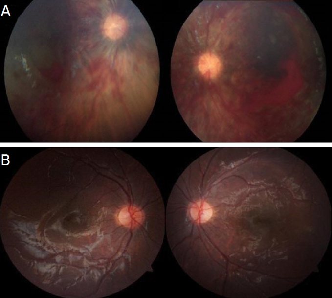
경미한 두부 외상력, 발작이나 의식저하로 본원 권역외상센터로 내원하여 학대뇌손상으로 확인되어 안과검사가 의뢰된 영유아는 총 14명으로 남아 7명, 여아 7명이었다. 의뢰된 나이는 평균 7.4개월(2-35개월)이었다. 모든 환아는 외상성 경막하출혈이 있었고, 두개골 골절이 1명, 팔뼈 골절이 2명에서 있었다(Fig. 1B, C). 이 가운데 9명(남아 5명, 여아 4명)에서 다발성 망막출혈이 있었다(Fig. 1D-I). 망막출혈이 있었던 9명의 평균 나이는 6개월(2-17개월)이었다. 망막출혈이 있었던 9명 중 8명(88.9%)이 뇌출혈 배액술이나 감압술과 같은 신경외과수술을 시행받았고, 망막출혈이 없었던 5명 중 1명(20.0%)이 뇌출혈 배액술을 시행받았다. 망막출혈이 있었던 환아 중 2명은 사망하고, 3명은 중증 발달지연, 1명은 언어지연과 같은 경증 발달지연이 있었고, 1명만 연령에 맞는 발달을 보였으며, 2명은 망막출혈이 흡수된 이후로는 경과 관찰되지 않았다(Table 1). 망막출혈이 없었던 5명의 환아 중에는 1명이 경증 발달지연이 있었다.
망막출혈은 출혈량 차이는 있어도 두 눈 모두에서 다발성으로 발생하였고, 첫 진단 후 평균 2.4개월(2-4개월, 2주 후 사망한 1명 제외)에 모두 흡수되어 망막수술이 필요한 경우는 없었다. 학대뇌손상에서 관찰될 수 있는 눈꺼풀 주변 푸른색 피부가 2명에서 관찰되었고(Fig. 1A), 뇌부종이 심한 2명에서는 동공이 확장된 채 고정되어 있었다. 휴대용 세극등현미경검사에서 결막출혈이나 결막하출혈, 전방출혈이 관찰되는 경우는 없었다. 망막출혈은 광범위하게 망막의 여러 층에서 발생하였고, 돔모양 망막출혈이 6명에서 관찰되었다. 황반부 주변 융기된 망막주름이 관찰된 1명은 출혈성 맥락막박리가 동반되어 있었다(Fig. 1H, case 2 in Table 1). 출혈이 흡수되면서 섬유질이 응고되어 하얀색 중심이 있는 망막출혈과 선상 망막출혈이 동시에 관찰되어 발생 시기가 다른 망막출혈이 함께 관찰되었다(Fig. 1I). 경과 관찰이 가능하였던 5명은 평균 28.2개월(9-51개월) 동안 경과 관찰하였으며 마지막 경과 관찰 시 안과적인 특이 소견이 없는 환아는 1명이었고, 두 눈 시신경유두창백 2명, 피질시각장애(cortical visual impairment) 1명, 한 눈 황반부 망막흉터와 시신경유두창백 1명이었다(Table 1, Fig. 2).

고 찰

학대뇌손상에서 경막하출혈은 영유아 머리를 앞뒤로 심하게 흔들어서 각가속-감속(angular acceleration-deceleration) 손상이 반복적으로 작용하여 대뇌피질과 정맥동을 연결하는 연결뇌정맥(bridge vessels)이 찢어져서 발생하는 것으로 알려져 있다.1,8 특히 영아는 머리가 몸에 비해 상대적으로 크고 목근육이 약해서 더 많은 손상을 받을 수 있다. 이와 같은 기전으로 광범위하게 망막의 여러 층에서 다량의 출혈이 나타날 수 있는데, 3세 미만 영유아에서 이러한 두개내출혈과 망막출혈이 함께 있으면 학대뇌손상의 예측도가 71%에 이른다.11 영유아에서는 유리체-망막부착이 견고하여 망막내경계막이 유리체에 부착된 채 망막출혈 주변 망막이 융기되어 하얀 테두리를 가지는 돔모양 망막출혈이 특징적으로 발생한다.1,3,12,14 유리체-망막부착이 견고한 황반부 주변으로 융기된 망막주름이 생기거나, 강한 유착이 있는 주변부 망막, 큰 망막혈관 주변, 황반부 주변으로 망막출혈이 조금 더 집중된다. 그리고 강한 유리체-망막부착으로 망막박리보다 망막층간분리가 흔히 발생한다.1,12,14,15 종종 발생 시기가 다른 망막출혈이 함께 있을 수 있어서 섬유소가 응집된 하얀색 중심이 있는 흡수 중인 출혈과 최근에 발생한 선홍색 출혈이 동시에 관찰될 수 있다.1,12,14 본 연구에 포함된 환아에서도 평균 생후 6.0개월에 황반부 주변 융기된 망막주름과 광범위한 다량의 망막출혈, 돔모양 망막출혈, 큰 망막혈관 주변에 집중된 망막출혈, 발생 시기가 다른 망막출혈이 함께 관찰되었다. 특히 황반부 주변 융기된 망막주름을 동반한 경우 사망률이 높은 것으로 알려져 있는데,1,3,12,14 본 연구에서도 그러한 융기된 망막주름이 관찰된 환아(case 2)는 사망하였다.
영유아에서 교통사고나 낙상사고로 인한 망막출혈은 출혈량이 상대적으로 적고 한 눈에 국한되어 발생하는 경우가 더 흔한 것으로 알려져 있다.1,6,7 출생 시 발생한 망막출혈은 대개 출생 후 2주 이내, 적어도 6주 이내에 모두 흡수된다.1,3,7 지주막하출혈, 뇌압상승이나 심폐소생술 때 흉부 압력 상승으로 인한 안내출혈은 시신경유두가 중심인 출혈이 발생한다.1,3,7 본 연구에 포함된 가장 어린 환아도 생후 10주였고, 심폐소생술을 시행받은 환아(case 1)를 포함한 모두 환아에서 시신경유두 주변보다는 광범위한 망막에서 많은 양의 망막출혈이 관찰되어 학대뇌손상에 의한 것으로 생각된다.
본 연구에서 학대뇌손상으로 안과에 의뢰된 영유아 14명 가운데 9명(64.3%)에서 망막출혈이 확인되었다. 학대뇌손상의 50-85%에서 망막출혈이 관찰된다는 이전 보고들과 일치하지만,1 실제로는 더 많은 환아에서 망막출혈이 있었을 수 있다. 영유아에서는 표면적이거나 크기가 작은 점상망막출혈은 그 수가 많더라도 24시간 이내에 흡수될 수 있어 빠른 안저검사가 필요한 것으로 알려져 있다.1,3,11 안과로 의뢰한 1-2일 이내 안과검사를 하였지만, 신경학적 응급 상황으로 인해 외상 후 평균 3.4일이 경과한 상태였기 때문에 적은 양의 망막출혈은 이미 흡수된 상태였을 수 있다. 더욱이 의뢰된 모든 환아는 경막하출혈을 포함한 외상성 두개출혈이 확인된 상태였는데, 머리외상과 다발성 망막출혈이 있다면 단순 망막출혈에 비해 학대뇌손상일 위험도가 14.7배 증가하는 것으로 알려져 있다.9
학대뇌손상에 의한 망막출혈이 있었던 경우 안과적 합병증이 흔히 동반된다.16-18 적어도 한 눈에 연령에 비해 시력 저하가 있는 경우가 46%, 사시 43%, 약시 40%에서 관찰되었으며, 시신경유두창백이 13%, 피질시각장애도 19%에서 있었다.16 본 연구에서도 망막출혈이 있었던 9명의 환아 가운데 정상 시력인 환아는 단 1명뿐이었다. 망막출혈이 있었던 환아 중 88.9%가 신경외과수술이 필요할 정도로 위중한 환아가 본 연구에 많이 포함되어 이전 보고보다 시력 예후가 더 좋지 않았던 것으로 생각된다.
본 연구는 후향적으로 진행되어 몇 가지 제한점이 있다. 국내 학대뇌손상 유병률이 아직 보고되지 않았지만, 미국의 연간 유병률(15-25/100,000명)을 감안하면1 비록 본원이 국내에서 가장 큰 권역외상센터이지만 확인되지 못한 학대뇌손상 환아가 더 많을 것으로 추정된다. 또한 환아 2명은 망막출혈이 흡수된 후 보호자가 더 이상 경과 관찰을 위해 내원하지 않았고, 퇴원 후 보육시설에서 양육된 환아 2명은 타지역으로 이관되어 지속적인 경과 관찰이 어려웠다. 휴대용 빛간섭단층촬영 장비가 없어서 출혈성 망막층간분리와 같은 망막-유리체유착에 의한 특징적인 병변들을 확인하지 못하였다.14 하지만, 본 연구는 단일권역외상센터에서 12년 동안 확인된 학대뇌손상 환아의 안과 소견을 기술한 국내 최초 보고라는 의미가 있다.
아동학대가 증가하고 있고, 더욱이 COVID-19기간을 지나면서 빠르게 증가하고 있다고 한다.19 아동학대가 의심되는 상황에서 철저한 안과검사가 필요하고, 3세 이하, 특히 생후 6-7개월 영유아가 뚜렷한 외상력 없이 다량의 망막출혈이 다발성으로 두 눈에서 확인되면 학대뇌손상을 감별 질환에 포함하여야 하겠다.

NOTES

Conflicts of Interest

The authors have no conflicts to disclose.

Acknowledgments

This work was supported by the National Research Foundation of Korea (NRF) grant funded by the Korea government (MSIT) (No. 2022R1F1A10 74170).

Figure 1.
Representative cases of abusive head trauma corroborated by pertinent findings. (A) External photographs with pale blue periocular skin (case 1, case 7). (B) X-ray with long bone fractures of the right radius and ulnar (white arrows, case 2). (C) Coronal view of brain computed tomography with subdural hematomas at different stages (arrow heads, case 3). (D-I) Fundus photographs with numerous extensive multilayered retinal hemorrhages including dome shape hemorrhages (cases 1-3, 5-7). (H) Perimacular folds as white retinal ridges surrounding the macula (yellow arrows, case 2), which is a well-established finding associated with fatal abusive head trauma. (I) Hemorrhages with white centers of clumped fibrin and fresh retinal hemorrhages supporting different attacks (case 6). The patient's legal guardians for (A) provided written informed consent for publication of the clinical images.
jkos-2024-65-3-235f1.jpg
Figure 2.
Fundus photographs of case 5. (A) Multiple retinal hemorrhages, which are more extensive in the left eye at age of 9 months. (B) Decreased foveal reflex and mild disc pallor in the left eye at age of 3.4 years (41 months).
jkos-2024-65-3-235f2.jpg
Table 1.
Findings for 9 cases of abusive head trauma with multiple retinal hemorrhages
Case Sex AI (mo) Ophthalmic findings (period of resorption of RH) General findings AF (mo) Ophthalmic sequelae Neurological sequelae
1 M 2 RH (2 mo) SDH, bilateral frontoparietal convexity 4 NLP, mild disc pallor (OU) Died
2 F 17 RH, perimacular folds SDH, left cerebral convexity 17 NLP (OU) Died
Fixed dilated pupil Fracture, right radius and ulnar shaft
3 M 3 RH (2 mo) SDH, right cerebral convexity 5 Follow-up loss Follow-up loss
Fracture, right distal radius
4 M 2 RH (2 mo) SDH, bilateral frontoparietal convexity 23 Left gaze preference Global developmental delay
Fixed dilated pupil Fracture, bilateral parietal bone No following (OU)
5 M 9 RH (4 mo) SDH, left cerebral convexity and tentorium 41 Amblyopia with mild disc pallor and foveal scar (OS) Speech delay
6 M 2 RH (2 mo) SDH, bilateral frontotemporal convexity 53 Normal vision (1.0/1.0) -
7 F 10 RH (3 mo) SDH, bilateral cerebral convexity and posterior fossa 19 Mild disc pallor (OU) Global developmental delay
Fracture, right parietal and occipital bones
8 F 2 RH (3 mo) SDH, posterior falx and right parietal convexity and tentorium 5 Follow-up loss Follow-up loss
9 F 7 RH (1 mo) SDH, left frontotemporal convexity 35 Right exotropia Global developmental delay
Disc pallor (OU)

AI (mo) = age at injury in months; RH = retinal hemorrhage; AF (mo) = age at last follow-up in months; M = male; SDH = subdural haemorrhage; NLP = no perception of light; OU = oculi uterque; F = female; OS = oculus sinister.

REFERENCES

1) Levin AV. Retinal hemorrhage in abusive head trauma. Pediatrics 2010;126:961-70.
crossref pmid pdf
2) Pierre-Kahn V, Roche O, Dureau P, et al. Ophthalmologic findings in suspected child abuse victims with subdural hematomas. Ophthalmology 2003;110:1718-23.
crossref pmid
3) Maguire SA, Watts PO, Shaw AD, et al. Retinal haemorrhages and related findings in abusive and non-abusive head trauma: a systematic review. Eye (Lond) 2013;27:28-36.
crossref pmid pdf
4) Ng WS, Watts P, Lawson Z, et al. Development and validation of a standardized tool for reporting retinal findings in abusive head trauma. Am J Ophthalmol 2012;154:333-9.e5.
crossref pmid
5) Oliva A, Grassi S, Cazzato F, et al. The role of retinal imaging in the management of abusive head trauma cases. Int J Legal Med 2022;136:1009-16.
crossref pmid pdf
6) Vincent AL, Kelly P. Retinal haemorrhages in inflicted traumatic brain injury: the ophthalmologist in court. Clin Exp Ophthalmol 2010;38:521-32.
crossref pmid
7) Harris CK, Stagner AM. The eyes have it: how critical are ophthalmic findings to the diagnosis of pediatric abusive head trauma? Semin Ophthalmol 2023;38:3-8.
crossref pmid
8) Maguire S, Pickerd N, Farewell D, et al. Which clinical features distinguish inflicted from non-inflicted brain injury? A systematic review. Arch Dis Child 2009;94:860-7.
crossref pmid
9) Weiss R, He CH, Khan S, et al. Ocular injuries in pediatric patients admitted with abusive head trauma. Pediatr Neurol 2022;127:11-8.
crossref pmid
10) Jeon TY, Yoo SY, Eo H, Kim JH. Diagnostic imaging for physical abuse in children. J Korean Soc Radiol 2020;81:832-51.
crossref pdf
11) Bhardwaj G, Jacobs MB, Martin FJ, et al. Photographic assessment of retinal hemorrhages in infant head injury: the childhood hemorrhagic retinopathy study. J AAPOS 2017;21:28-33.e2.
crossref pmid
12) Breazzano MP, Unkrich KH, Barker-Griffith AE. Clinicopathological findings in abusive head trauma: analysis of 110 infant autopsy eyes. Am J Ophthalmol 2014;158:1146-54.e2.
crossref pmid
13) Kim YH, Choi SY, Lee JS, et al. Smartphone fundus photography in an infant with abusive head trauma. J Korean Ophthalmol Soc 2017;58:1313-6.
crossref pdf
14) Matschke J, Püschel K, Glatzel M. Ocular pathology in shaken baby syndrome and other forms of infantile non-accidental head injury. Int J Legal Med 2009;123:189-97.
crossref pmid pdf
15) Muni RH, Kohly RP, Sohn EH, Lee TC. Hand-held spectral domain optical coherence tomography finding in shaken-baby syndrome. Retina 2010;30:S45-50.
crossref pmid
16) Weldy E, Shimoda A, Patnaik J, et al. Long-term visual outcomes following abusive head trauma with retinal hemorrhage. J AAPOS 2019;23:329.e1-4.
crossref pmid
17) Kelly JP, Feldman KW, Wright JN, et al. Pediatric abusive head trauma: visual outcomes, evoked potentials, diffusion tensor imaging, and relationships to retinal hemorrhages. Doc Ophthalmol 2023;147:1-14.
crossref pmid pmc pdf
18) Chong CF, Misra SL, Escardo-Paton JA, Dai S. Predictors of long-term neurological outcomes in non-accidental head injury. Eye (Lond) 2018;32:608-14.
crossref pmid pdf
19) Lãzãrescu AM, Benichi S, Blauwblomme T, et al. Abusive head trauma in infants during the COVID-19 pandemic in the Paris metropolitan area. JAMA Netw Open 2022;5:e2226182.
crossref pmid pmc

Biography

우태경 / Taekyeong Woo
Department of Ophthalmology, Ajou University School of Medicine
TOOLS
METRICS Graph View
  • 0 Crossref
  •  0 Scopus
  • 403 View
  • 45 Download
Related articles


ABOUT
BROWSE ARTICLES
EDITORIAL POLICY
FOR CONTRIBUTORS
Editorial Office
SKY 1004 Building #701
50-1 Jungnim-ro, Jung-gu, Seoul 04508, Korea
Tel: +82-2-583-6520    Fax: +82-2-583-6521    E-mail: kos08@ophthalmology.org                

Copyright © 2024 by Korean Ophthalmological Society.

Developed in M2PI

Close layer
prev next